. 2015 Aug;95(8):951-61.
doi: 10.1038/labinvest.2015.62. Epub 2015 Jun 1.

The hepatocyte growth factor receptor as a potential therapeutic target for dedifferentiated liposarcoma

Affiliations

The hepatocyte growth factor receptor as a potential therapeutic target for dedifferentiated liposarcoma

Kate Lynn J Bill et al. Lab Invest. 2015 Aug.

Abstract

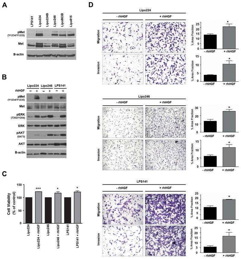
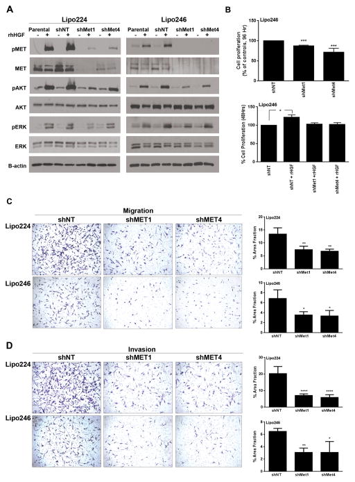
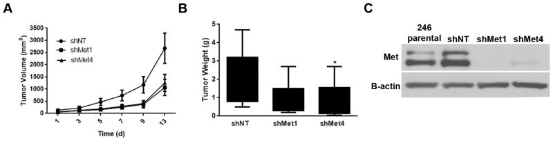
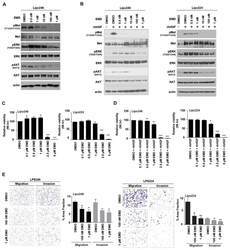
Dedifferentiated liposarcomas (DDLPS) are highly resistant to conventional chemo- and radiotherapies, with surgical resection remaining the classic treatment strategy; therefore, there is a pressing need for novel anti-DDLPS-targeted chemotherapeutics. Hepatocyte growth factor receptor (Met) expression is elevated in DDLPS, but the functional role of Met signaling in this disease is not known. We found that the in vitro stimulation of DDLPS cells with hepatocyte growth factor (HGF) elevated the degree of PI3K/AKT and MAPK pathway signaling, and that pro-tumorigenic phenotypes such as cell proliferation, invasion, and migration were significantly enhanced. Conversely, Met knockdown using shRNA-mediated interference decreased HGF-induced Met signaling, the invasive and migratory nature of DDLPS cells in vitro, and the tumorigenicity of DDLPS cells in vivo. These data strongly support the role for Met as a DDLPS therapeutic target. To that end, using EMD1214063, an ATP-competitive kinase inhibitor that targets Met more specifically than other kinases, inhibited Met-dependent signaling, reduced the oncogenicity of DDLPS cells in vitro, and significantly increased the survival of nude mice bearing subcutaneous DDLPS xenografts. These findings support further investigations of HGF-induced Met signaling inhibition in DDLPS, as a potential strategy to enhance clinical outcomes for this disease.


Conflict of interest statement

Disclosure/Duality of Interest:

The authors declare no conflict of interest.

Figures

Figure 1
Figure 1. HGF-mediated Met activation enhances oncogenic signaling and phenotypes of DDLPS cells in vitro
(A) Western blot analysis of activated Met in several DDLPS cell lines (10% FBS media). (B) Western blots monitored HGF-stimulated Met, and downstream Erk1/2 and AKT activation in DDLPS cells. Lipo224, Lipo246, and LPS141 cells were serum-starved overnight and stimulated with rhHGF (50 ng/mL) for 20 minutes. (C) MTS assays measured cell proliferation of 24 hr serum-starved DDLPS cells over a 48 hr period with or without rhHGF (50 ng/mL) stimulation (n=3 ± SEM; t-test: *=P<0.05, ***=P<0.0001; samples were analyzed at least in duplicate per experiment). (D) Modified Boyden chamber assays measured the invasion and migration capabilities of DDLPS cells with or without the use of rhHGF (50 ng/mL) as a chemoattractant. (Images are representative, and graphs represent n=3 experiments ± SEM; t-test: *=P<0.05, **=P<0.005, ***=P<0.0001; samples were analyzed at least in duplicate per experiment).
Figure 2
Figure 2. Met knockdown decreases PI3K and MAPK pathway signaling, and the proliferative, invasive, and migratory phenotypes of DDLPS cells in vitro
(A) Left panel: Western blot analyses of downstream signaling in Met knockdown Lipo224 cells that were serum-starved and then stimulated with 50 ng/mL rhHGF for 15 minutes (− represents no rhHGF stimulation; + represents cells that were stimulated with rhHGF). Right panel: Western analysis was performed as in the left panel, however this panel evaluated Lipo246 cells with Met knockdown and rhHGF stimulation. (B) Upper Panel: MTS assays measured the proliferative capabilities of shNT, shMet1, and shMet4 polyclonal populations after 96 hours in culture in Lipo246 Met knockdown cells (10%-FBS supplemented media; n=3 experiments ± SEM; t-test: *=P<0.05, ***=P<0.0001; samples were analyzed at least in duplicate per experiment). Lower Panel: 50 ng/mL rhHGF stimulated Lipo246 DDLPS cells with Met knockdown were serum-starved for 24 h prior to stimulation with 50 ng/mL rhHGF for 48 h. MTS assays measured cell proliferation (n=3 experiments ± SEM; t-test: *=P<0.05, **=P<0.005, ***=P<0.0001; samples were analyzed at least in duplicate per experiment). Lipo224 and Lipo246 shMet cells exhibit a statistically significant decrease in migration (C) and invasion (D) when compared with shNT cells (rhHGF was used as a chemoattractant; n=3 experiments ± SEM; t-test: *=P<0.05, **=P<0.005, ***=P<0.0001; samples were analyzed at least in duplicate per experiment).
Figure 3
Figure 3. Met knockdown decreases tumorigenicity of DDLPS cells in vivo
(A) Lipo246 cells expressing a non-targeting construct (shNT), or two different shMet targeting constructs were injected subcutaneously, and the tumor volume measured over time. Met KD xenografts had an overall slower growth rate and a decreased tumor weight (B) at termination as compared to control shNT tumor bearing control mice. (Mean tumor volume at 12 days for each group of mice was recorded ± SEM; t-test: *=P<0.05) (C) Western blot confirmed Met KD in xenograft tumors.
Figure 4
Figure 4. Decreased oncogenic signaling and aggressiveness of DDLPS cells with Met inhibition in vitro
(A) Western blot analysis monitored decreasing Met, AKT, and Erk1/2 activity levels in Lipo246 cells with increasing concentrations of EMD1214063 (4 h; 10%-FBS containing media). (B) Left Panel; Lipo246 cells. Right Panel; Lipo224 cells: Serum-starved cells were pretreated with increasing concentrations of EMD1214063 followed by rhHGF stimulation for 20 min. Western blot analyses revealed that Met, AKT, and ERK1/2 signaling decreased in a dose-dependent manner. (C) Left Panel: Lipo246; Right panel: Lipo224. MTS assays measured proliferation rates as a consequence of increasing EMD1214063 treatment (n=3 experiments ± SEM; t-test: *=P<0.05, **=P<0.005, ***=P<0.0001; samples were analyzed at least in duplicate per experiment). (D) Serum starved Lipo246 (Left Panel) and Lipo224 (Right Panel) were simultaneously treated with increasing concentrations of EMD1214063 and rhHGF (50 ng/mL) for 96 h. MTS assays revealed that proliferation rates decreased with increasing concentrations of EMD1214063 (n=3 experiments ± SEM; t-test: *=P<0.05, **=P<0.005, ***=P<0.0001; samples were analyzed at least in duplicate per experiment). (E) Left Panel: Lipo246 cells were either treated with DMSO, 100 nM, or 1 μM EMD1214063 for 24 h, and migration and invasion results recorded (3 images per well; at least 2 replicates per experiment; n=3 experiments ± SEM; t-test: *=P<0.05). Right Panel: Lipo224 cells were also either treated with DMSO, 100 nM, or 1 μM EMD1214063 for 24 h (3 images per well; at least 2 replicates per experiment; n=3 experiments ± SEM; t-test: *=P<0.05, **=P<0.005).
Figure 5
Figure 5. Met kinase inhibition decreases the tumorigenicity of Lipo246 xenografts in vivo
(A) EMD1214063 treatment of Lipo246 xenografts decreased tumor volume and (B) tumor weight significantly within 17 days. (C) Western blots monitored whether EMD1214063 inhibited Met Y1234/Y1235 phosphorylation in EMD1214063 treated xenografts compared with vehicle control treated xenografts.

Similar articles

Cited by

References

    1. Lahat G, Zhu Q-S, Huang K-L, Wang S, Bolshakov S, Liu J, et al. Vimentin is a novel anti-cancer therapeutic target; insights from in vitro and in vivo mice xenograft studies. In: Bauer JA, editor. PLoS One. 4. Vol. 5. Public Library of Science; 2010. Jan, p. e10105. - PMC - PubMed
    1. Taylor BS, DeCarolis PL, Angeles CV, Brenet F, Schultz N, Antonescu CR, et al. Frequent alterations and epigenetic silencing of differentiation pathway genes in structurally rearranged liposarcomas. Cancer Discov. 2011 Dec;1(7):587–97. - PMC - PubMed
    1. Zhang P, Bill K, Liu J, Young E, Peng T, Bolshakov S, et al. MiR-155 is a liposarcoma oncogene that targets casein kinase-1α and enhances β-catenin signaling. Cancer Res. 2012 Apr 1;72(7):1751–62. - PMC - PubMed
    1. Hoffman A, Lazar AJ, Pollock RE, Lev D. Drug Resist Updat. 1. Vol. 14. Elsevier Ltd; 2011. Feb, New frontiers in the treatment of liposarcoma, a therapeutically resistant malignant cohort; pp. 52–66. - PubMed
    1. Singer S, Socci ND, Ambrosini G, Sambol E, Decarolis P, Wu Y, et al. Gene expression profiling of liposarcoma identifies distinct biological types/subtypes and potential therapeutic targets in well-differentiated and dedifferentiated liposarcoma. Cancer Res. 2007 Jul 15;67(14):6626–36. - PubMed

Publication types

MeSH terms