

Brain activation in response to overt and covert fear and happy faces in women with borderline personality disorder
- PMID: 26007149
- PMCID: PMC5549680
- DOI: 10.1007/s11682-015-9406-4
Brain activation in response to overt and covert fear and happy faces in women with borderline personality disorder
Abstract
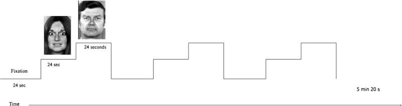
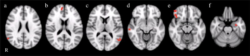
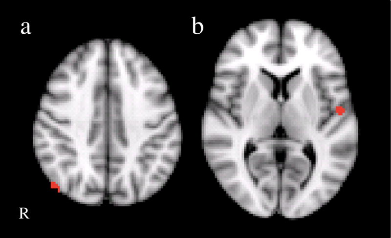
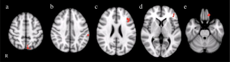
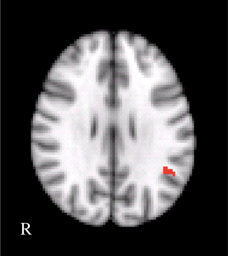
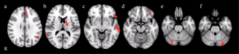
Borderline personality disorder (BPD) is a serious condition involving emotion dysregulation. Past research has identified BPD-associated differences within fronto-limbic circuitry during conditions of processing negative emotion. Functional magnetic resonance imaging (fMRI) paradigms that incorporate overt and covert (masked) presentations of emotional stimuli can provide complementary information about neural systems underlying emotion processing (e.g., both slow [overt] and fast [covert; automatic] processing pathways). This study examined brain activation during processing of overt and covert presentations of emotional faces in 12 women with BPD and 12 age-matched healthy controls. To assess a range of emotional valence and arousal, we examined responses to fear, happy and neutral expressions. All participants underwent an fMRI scanning session in which participants passively viewed emotional faces. Scanning sessions consisted of 5 runs including: (1) Overt Fear (OF) versus Neutral (N), (2) Covert Fear (CF) versus Covert Neutral (CN), (3) Overt Happy (OH) versus N, (4) Covert Happy (CH) versus CN, and (5) N versus fixation. We compared whole-brain activation between groups for each run. In response to overt fear, BPD patients showed greater activation both in left amygdala and in several frontal cortical regions. There were no significant differences in brain activation in response to overt happy faces. In response to covert fear and covert happy stimuli, the BPD group also showed greater activation than controls in several regions including frontal and temporal cortical regions, as well as cerebellum and thalamus. These findings add to prior reports suggesting increased amygdala activation in BPD, but we found this only in the overt fear versus fixation condition. In this sample, BPD patients showed hyper-activation, rather than hypo-activation, of cortical regulatory regions during overt fear. Enhanced cortical recruitment in response to covert fear and happy faces in BPD could reflect a more extended response system in which stimuli that typically only activate automatic pathways are additionally tapping into cortical regulatory systems. The observation of this pattern both in response to fear and in response to happy presentations suggests that the effect of arousal may be as or more impactful than the effect of emotional valence.
Keywords: Amygdala; Backward masking; Borderline personality disorder; Covert; Emotion; Overt; fMRI.
Conflict of interest statement
Figures
Similar articles
-
Fronto-limbic dysfunction in response to facial emotion in borderline personality disorder: an event-related fMRI study.Psychiatry Res. 2007 Aug 15;155(3):231-43. doi: 10.1016/j.pscychresns.2007.03.006. Epub 2007 Jul 2. Psychiatry Res. 2007. PMID: 17601709 Free PMC article.
-
Amygdala functional connectivity in young women with borderline personality disorder.Brain Connect. 2011;1(1):61-71. doi: 10.1089/brain.2010.0001. Brain Connect. 2011. PMID: 22432955 Free PMC article.
-
Differential neural responses to overt and covert presentations of facial expressions of fear and disgust.Neuroimage. 2004 Apr;21(4):1484-96. doi: 10.1016/j.neuroimage.2003.12.013. Neuroimage. 2004. PMID: 15050573
-
Functional atlas of emotional faces processing: a voxel-based meta-analysis of 105 functional magnetic resonance imaging studies.J Psychiatry Neurosci. 2009 Nov;34(6):418-32. J Psychiatry Neurosci. 2009. PMID: 19949718 Free PMC article. Review.
-
Defining the neurocircuitry of borderline personality disorder: functional neuroimaging approaches.Dev Psychopathol. 2005 Fall;17(4):1197-206. doi: 10.1017/s095457940505056x. Dev Psychopathol. 2005. PMID: 16613437 Review.
Cited by
-
The NIMH Research Domain Criteria (RDoC) Initiative and Its Implications for Research on Personality Disorder.Curr Psychiatry Rep. 2019 Apr 27;21(6):37. doi: 10.1007/s11920-019-1023-2. Curr Psychiatry Rep. 2019. PMID: 31030293 Review.
-
Brain Activation for Social Cognition and Emotion Processing Tasks in Borderline Personality Disorder: A Meta-Analysis of Neuroimaging Studies.Brain Sci. 2024 Apr 18;14(4):395. doi: 10.3390/brainsci14040395. Brain Sci. 2024. PMID: 38672044 Free PMC article. Review.
-
Development and validation of an emotional picture set of self-injury (EPSI) for borderline personality disorder: protocol for a validation study.BMJ Open. 2019 May 22;9(5):e027063. doi: 10.1136/bmjopen-2018-027063. BMJ Open. 2019. PMID: 31122985 Free PMC article.
-
Facial emotion processing in patients with borderline personality disorder as compared with healthy controls: an fMRI and ECG study.Borderline Personal Disord Emot Dysregul. 2024 Feb 16;11(1):4. doi: 10.1186/s40479-024-00245-4. Borderline Personal Disord Emot Dysregul. 2024. PMID: 38360712 Free PMC article.
-
Attentional bias during emotional processing: Behavioral and electrophysiological evidence from an Emotional Flanker Task.PLoS One. 2021 Apr 2;16(4):e0249407. doi: 10.1371/journal.pone.0249407. eCollection 2021. PLoS One. 2021. PMID: 33798215 Free PMC article.
References
-
- Beblo T, Driessen M, Mertens M, Wingenfeld K, Piefke M, Rullkoetter N, Silva-Saavedra A, Mensebach C, Reddemann L, Rau H, Markowitsch HJ, Wulff H, Lange W, Berea C, Ollech I, Woermann FG. Functional MRI correlates of the recall of unresolved life events in borderline personality disorder. Psychological Medicine. 2006;36:845–856. doi: 10.1017/S0033291706007227. - DOI - PubMed
-
- Birbaumer N, Grodd W, Diedrich O, Klose U, Erb M, Lotze M, Schneider F, Weiss U, Flor H. fMRI reveals amygdala activation to human faces in social phobics. Neuroreport. 1998;9:1223–1226. - PubMed
-
- Breiter HC, Etcoff NL, Whalen PJ, Kennedy WA, Rauch SL, Buckner RL, Strauss MM, Hyman SE, Rosen BR. Response and habituation of the human amygdala during visual processing of facial expression. Neuron. 1996;17:875–887. - PubMed
MeSH terms
Grants and funding
LinkOut - more resources
Full Text Sources
Other Literature Sources
Research Materials
Miscellaneous
