Review
. 2015 Jul;11(7):393-405.
doi: 10.1038/nrendo.2015.82. Epub 2015 May 26.

Perimenopause as a neurological transition state

Affiliations
Review

Perimenopause as a neurological transition state

Roberta D Brinton et al. Nat Rev Endocrinol. 2015 Jul.

Abstract

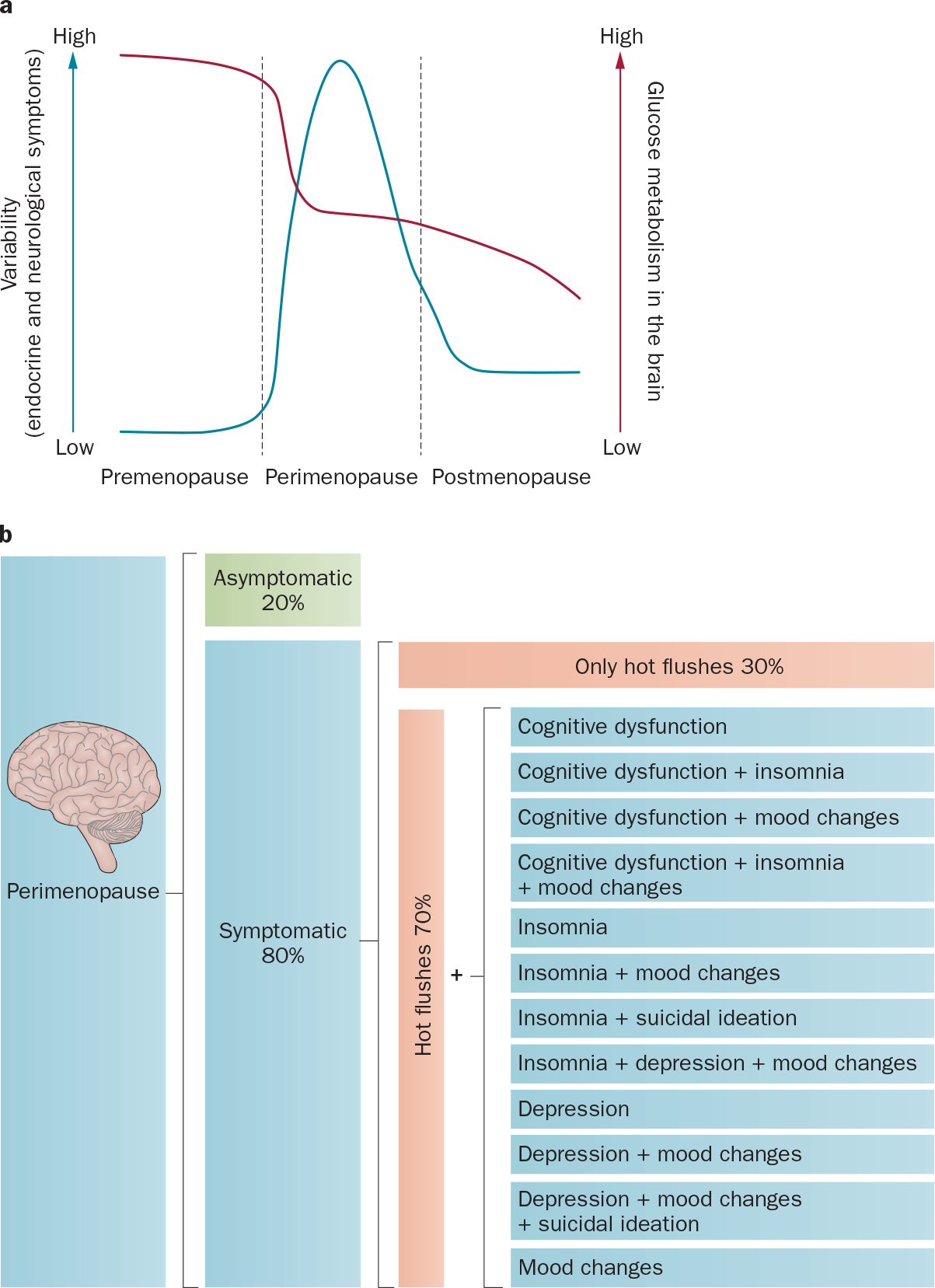
Perimenopause is a midlife transition state experienced by women that occurs in the context of a fully functioning neurological system and results in reproductive senescence. Although primarily viewed as a reproductive transition, the symptoms of perimenopause are largely neurological in nature. Neurological symptoms that emerge during perimenopause are indicative of disruption in multiple estrogen-regulated systems (including thermoregulation, sleep, circadian rhythms and sensory processing) and affect multiple domains of cognitive function. Estrogen is a master regulator that functions through a network of estrogen receptors to ensure that the brain effectively responds at rapid, intermediate and long timescales to regulate energy metabolism in the brain via coordinated signalling and transcriptional pathways. The estrogen receptor network becomes uncoupled from the bioenergetic system during the perimenopausal transition and, as a corollary, a hypometabolic state associated with neurological dysfunction can develop. For some women, this hypometabolic state might increase the risk of developing neurodegenerative diseases later in life. The perimenopausal transition might also represent a window of opportunity to prevent age-related neurological diseases. This Review considers the importance of neurological symptoms in perimenopause in the context of their relationship to the network of estrogen receptors that control metabolism in the brain.


Conflict of interest statement

Competing interests

The authors declare no competing interests.

Figures

Figure 1 |
Figure 1 |
Symptoms during perimenopause. Perimenopause is characterized by heightened variability in neurological symptoms, which is co-incident with a decline in glucose metabolism in the brain. a | Variability of symptoms in premenopause, perimenopause and postmenopause (blue line; left axis); glucose metabolism in the brain (red line; right axis). b | Diversity of neurological symptoms during perimenopause. The majority of women will experience neurological symptoms during perimenopause; however, a small proportion (~20%) of women will transition without symptoms.
Figure 2 |
Figure 2 |
Neurological functions affected by the perimenopausal transition. Brain regions and their corresponding functions provide a map of the neural circuits regulated by estrogen and a neurobiological basis for the array of symptoms that can emerge during perimenopause. Nuclear, membrane-associated and mitochondrial estrogen receptors are distributed within each of the neural circuits and can be present in both neurons and glial cells. While the complete distribution of ER-α and ER-β remains to be completely mapped in humans, in rats the location of these receptors is well documented. Dysregulation of estrogen signalling, either through changes in estrogen concentration or through modifications of estrogen receptor, will affect neural circuit activation and, thus, neurological function. Abbreviations: ER, estrogen receptor; GPER, G protein-coupled estrogen receptor.
Figure 3 |
Figure 3 |
Estrogen receptor network. The estrogen receptor network integrates cellular responses and functions in the brain. Estrogen binding and activation of the membrane-bound receptors, mER-α, mER-β and GPER, leads to activation of signalling pathways that regulate expression of early and intermediate response genes. Estrogen binding to the nuclear estrogen receptors, ER-α and ER-β, results in activation of transcriptional pathways that regulate expression of late response genes. Activation and translocation of ER-β to the mitochondria has been implicated in regulating expression of mitochondrial genes. Furthermore, estrogen can modulate transcriptional gene expression, activated by either rapid signalling or transcriptional pathways, via epigenetic regulation. This network of receptors enables the integration of signals across rapid, intermediate and late response pathways to coordinate a broad spectrum of cellular elements, including substrate transporters, metabolic enzymes and catalytic processes, which ultimately results in generation of energy to fuel neurological function. Abbreviations: ER, estrogen receptor; GPER, G-protein coupled estrogen receptor 1; mER, membrane estrogen receptor; mtER, mitochondrial estrogen receptor.
Figure 4 |
Figure 4 |
Estrogen-mediated regulation of the bioenergetic system. Estrogen signalling supports and sustains glucose metabolism in the brain by regulating expression of glucose transporters, which results in increased glucose uptake, and by stimulating glucose metabolism, mitochondrial oxidative phosphorylation and ATP generation—collectively referred to as aerobic glycolysis. Glucose (1) is the primary metabolic fuel for the brain. Estrogen regulates the bioenergetic system in brain the through the estrogen receptors, GPER, ER-α and ER-β, and their activation of PI3K and downstream Akt and MAPK–ERK signalling pathways. When the glucose pathway is compromised, for example, during starvation, acetyl-CoA can be generated from ketone bodies via ketogenesis in the liver and transported through the blood to the brain through monocarboxylate transporters (2) or from fatty acid via β-oxidation (3). During the perimenopausal transition, neuronal levels of glucose transporters decline, which is co-incident with the appearance of hypometabolism in the brain. The brain adapts to this decline in glucose availability by increasing reliance on ketone bodies as an alternative fuel to generate acetyl-CoA required for entry to the TCA cycle (4) and ultimately generation of ATP via complexes of the mitochondrial redox carriers (5). Initially, ketone bodies are derived from the periphery by lipid metabolism in the liver. Depletion of peripheral sources of ketone bodies can result in metabolism of brain-derived fatty acids to generate ketone bodies via β-oxidation in glia cells (3). Abbreviations: GP, glucose-6-phosphate; GPER, G protein-coupled estrogen receptor 1; ER-α, estrogen receptor α; ER-β, estrogen receptor β; HK, hexokinase; α-KGDH, α-ketoglutarate dehydrongenase, IGF-1, insulin growth factor-1; IRS, insulin receptor substrate; MCT, monocarboxylate transporter; PDH, pyruvate dehydrogenase; PI3K, phosphoinositide 3-kinase; SCOT, succinyl-CoA:3-ketoacid CoA transferase; SDH, succinate dehydrogenase; TCA, tricarboxylic acid cycle.

References

    1. Brinton RD in Brockelhurst’s Textbook of Geriatric Medicine and Gerontology (eds Fillit H, Rockwood K & Young J) 163–171 (Saunders, 2010).
    1. Butler L & Santoro N The reproductive endocrinology of the menopausal transition. Steroids 76, 627–635 (2011). - PMC - PubMed
    1. Harlow SD et al. Executive summary of the Stages of Reproductive Aging Workshop + 10: addressing the unfinished agenda of staging reproductive aging. Menopause 19, 387–395, (2012). - PMC - PubMed
    1. Greendale GA, Ishii S, Huang MH & Karlamangla AS Predicting the timeline to the final menstrual period: the study of women’s health across the nation. J. Clin. Endocrinol. Metab. 98, 1483–1491 (2013). - PMC - PubMed
    1. United States Census Bureau. World population by age and sex, [online], http://www.census.gov/population/international/data/worldpop/tool_popula... (2014).

Publication types