

Applications of two-dimensional infrared spectroscopy
- PMID: 26007625
- PMCID: PMC4500793
- DOI: 10.1039/c5an00558b
Applications of two-dimensional infrared spectroscopy
Abstract
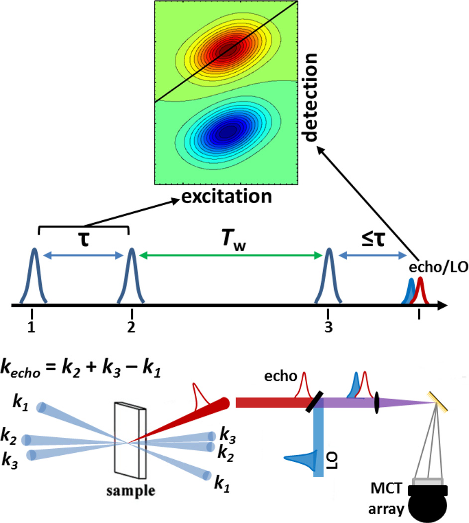
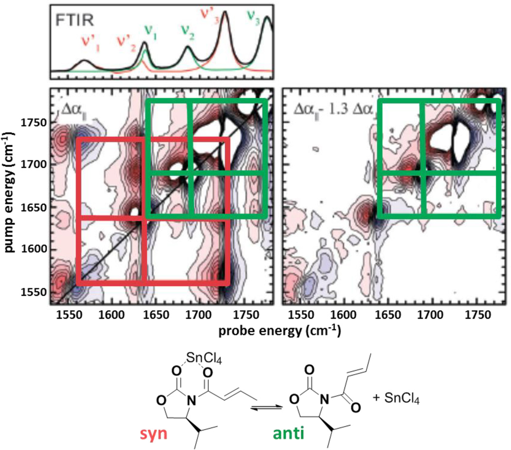
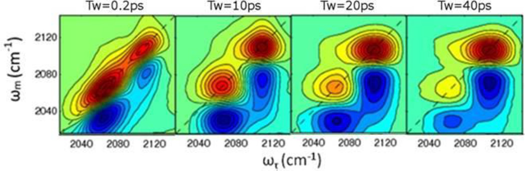
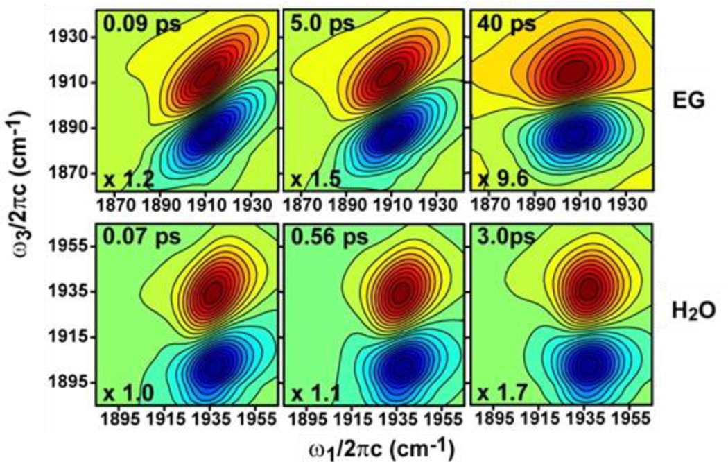
Two-dimensional infrared (2D IR) spectroscopy has recently emerged as a powerful tool with applications in many areas of scientific research. The inherent high time resolution coupled with bond-specific spatial resolution of IR spectroscopy enable direct characterization of rapidly interconverting species and fast processes, even in complex systems found in chemistry and biology. In this minireview, we briefly outline the fundamental principles and experimental procedures of 2D IR spectroscopy. Using illustrative example studies, we explain the important features of 2D IR spectra and their capability to elucidate molecular structure and dynamics. Primarily, this minireview aims to convey the scope and potential of 2D IR spectroscopy by highlighting select examples of recent applications including the use of innate or introduced vibrational probes for the study of nucleic acids, peptides/proteins, and materials.
Figures

Similar articles
-
Transparent window 2D IR spectroscopy of proteins.J Chem Phys. 2021 Jul 28;155(4):040903. doi: 10.1063/5.0052628. J Chem Phys. 2021. PMID: 34340394 Free PMC article. Review.
-
Ultrafast structural molecular dynamics investigated with 2D infrared spectroscopy methods.Top Curr Chem (Cham). 2017 Oct 25;375(6):86. doi: 10.1007/s41061-017-0172-1. Top Curr Chem (Cham). 2017. PMID: 29071445 Review.
-
Using 2D-IR Spectroscopy to Measure the Structure, Dynamics, and Intermolecular Interactions of Proteins in H2O.Acc Chem Res. 2024 Mar 5;57(5):685-692. doi: 10.1021/acs.accounts.3c00682. Epub 2024 Feb 16. Acc Chem Res. 2024. PMID: 38364823 Free PMC article. Review.
-
Protein dynamics studied with ultrafast two-dimensional infrared vibrational echo spectroscopy.Acc Chem Res. 2012 Nov 20;45(11):1866-74. doi: 10.1021/ar200275k. Epub 2012 Mar 20. Acc Chem Res. 2012. PMID: 22433178 Free PMC article.
-
Protein Dynamics by Two-Dimensional Infrared Spectroscopy.Annu Rev Anal Chem (Palo Alto Calif). 2021 Jul 27;14(1):299-321. doi: 10.1146/annurev-anchem-091520-091009. Annu Rev Anal Chem (Palo Alto Calif). 2021. PMID: 34314221 Free PMC article.
Cited by
-
Intermolecular Vibration Energy Transfer Process in Two CL-20-Based Cocrystals Theoretically Revealed by Two-Dimensional Infrared Spectra.Molecules. 2022 Mar 26;27(7):2153. doi: 10.3390/molecules27072153. Molecules. 2022. PMID: 35408551 Free PMC article.
-
Extracting accurate infrared lineshapes from weak vibrational probes at low concentrations.MethodsX. 2023 Aug 1;11:102309. doi: 10.1016/j.mex.2023.102309. eCollection 2023 Dec. MethodsX. 2023. PMID: 37577166 Free PMC article.
-
Transparent window 2D IR spectroscopy of proteins.J Chem Phys. 2021 Jul 28;155(4):040903. doi: 10.1063/5.0052628. J Chem Phys. 2021. PMID: 34340394 Free PMC article. Review.
-
Dynamics underlying hydroxylation selectivity of cytochrome P450cam.Biophys J. 2021 Mar 2;120(5):912-923. doi: 10.1016/j.bpj.2021.01.027. Epub 2021 Feb 3. Biophys J. 2021. PMID: 33545101 Free PMC article.
-
Biomolecular infrared spectroscopy: making time for dynamics.Chem Sci. 2023 Nov 28;15(2):414-430. doi: 10.1039/d3sc05223k. eCollection 2024 Jan 3. Chem Sci. 2023. PMID: 38179520 Free PMC article. Review.
References
-
- Zheng J, Kwak K, Fayer MD. Acc. Chem. Res. 2007;40:75–83. - PubMed
-
- Park S, Kwak K, Fayer MD. Laser Phys. Lett. 2007;4:704–718.
-
- Mukamel S. Principles of Nonlinear Optics and Spectroscopy. New York: Oxford University Press; 1995.
-
- Hamm P, Zanni M. Concepts and Methods of 2D Infrared Spectroscopy. Cambridge University Press; 2011.
Publication types
MeSH terms
Substances
Grants and funding
LinkOut - more resources
Full Text Sources
Other Literature Sources