. 2015 Oct:82:12-21.
doi: 10.1016/j.nbd.2015.05.010. Epub 2015 May 22.

The deubiquitinase ataxin-3 requires Rad23 and DnaJ-1 for its neuroprotective role in Drosophila melanogaster

Affiliations

The deubiquitinase ataxin-3 requires Rad23 and DnaJ-1 for its neuroprotective role in Drosophila melanogaster

Wei-Ling Tsou et al. Neurobiol Dis. 2015 Oct.

Abstract

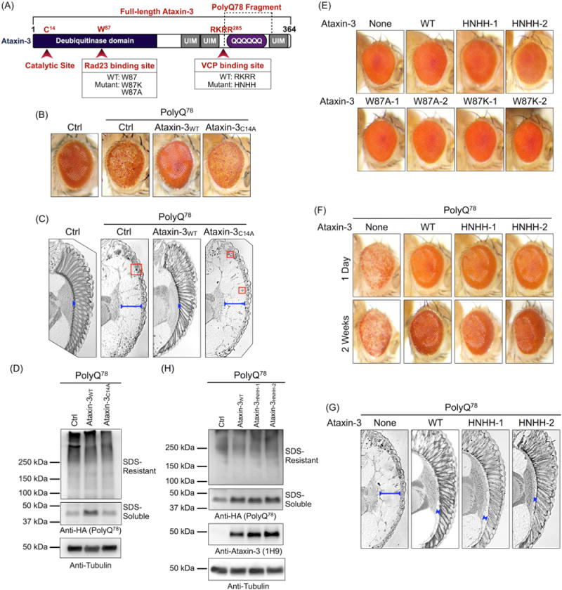
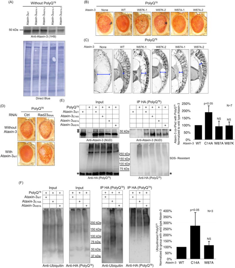
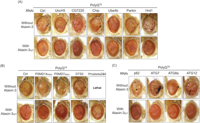
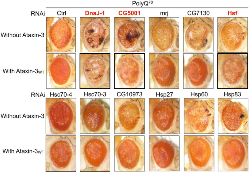
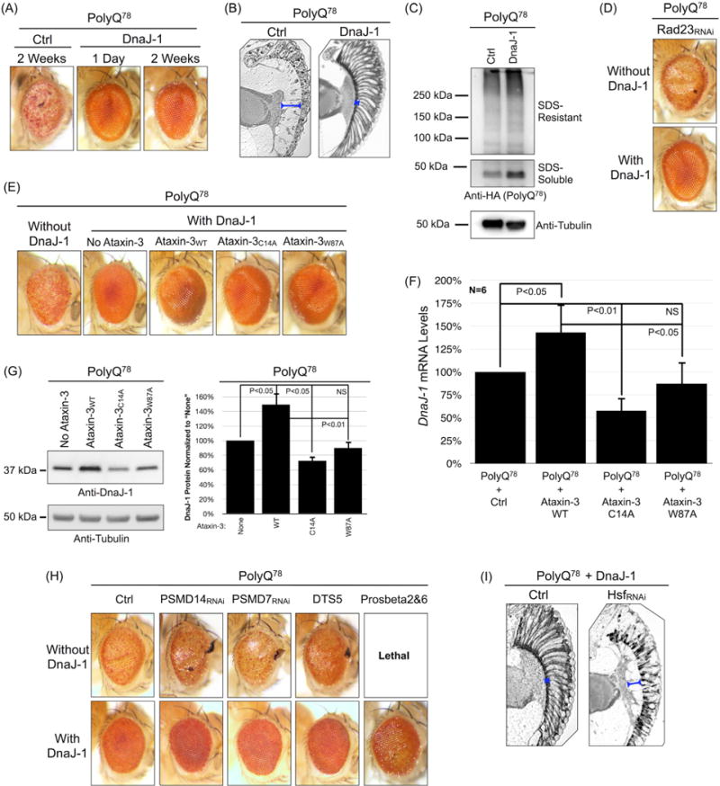
Ataxin-3 is a deubiquitinase and polyglutamine (polyQ) disease protein with a protective role in Drosophila melanogaster models of neurodegeneration. In the fruit fly, wild-type ataxin-3 suppresses toxicity from several polyQ disease proteins, including a pathogenic version of itself that causes spinocerebellar ataxia type 3 and pathogenic huntingtin, which causes Huntington's disease. The molecular partners of ataxin-3 in this protective function are unclear. Here, we report that ataxin-3 requires its direct interaction with the ubiquitin-binding and proteasome-associated protein, Rad23 (known as hHR23A/B in mammals) in order to suppress toxicity from polyQ species in Drosophila. According to additional studies, ataxin-3 does not rely on autophagy or the proteasome to suppress polyQ-dependent toxicity in fly eyes. Instead this deubiquitinase, through its interaction with Rad23, leads to increased protein levels of the co-chaperone DnaJ-1 and depends on it to protect against degeneration. Through DnaJ-1, our data connect ataxin-3 and Rad23 to protective processes involved with protein folding rather than increased turnover of toxic polyQ species.

Keywords: Ataxin-3; Chaperone; Deubiquitinase; Drosophila; Machado–Joseph disease; Polyglutamine; Ubiquitin.


Conflict of interest statement

Conflict of interest statement

The authors declare that they have no competing interests.

Figures

Fig. 1
Fig. 1
The VCP-ataxin-3 interaction is not necessary for protection against polyQ78 in Drosophila eyes. A) Diagram of the domain composition of the wild-type ataxin-3 protein. The N-terminal portion contains the catalytic domain. The catalytic cysteine is at position 14. At position 87 is a tryptophan residue that is important for the direct binding of ataxin-3 to Rad23 (Nicastro et al., 2005, 2009, 2010). Following the catalytic domain are ubiquitin-interacting motifs (UIMs) and the polyglutamine (polyQ) region, which causes SCA3 when expanded. Preceding the polyQ tract is the VCP-binding site. Highlighted with dashed lines is the part of ataxin-3 that was utilized to make the polyQ78 model of degeneration in Drosophila using an isoform of ataxin-3 that does not have the third UIM (Warrick et al., 1998). This construct comprises the polyQ fragment of ataxin-3 with a few amino acids preceding it without a functional VCP-binding area (Boeddrich et al., 2006; Warrick et al., 1998) and also includes part of the region following the polyQ tract without the third UIM. The specific mutations used for the Rad23-binding site and the VCP-binding site are noted below the diagram. B, C) External photos (B) and histological sections (C) of fly eyes expressing UAS-polyQ78 through the gmr-Gal4 driver in the absence (Ctrl) or presence of UAS-ataxin-3 wild-type (WT) or catalytically inactive (C14A). All flies were heterozygous for all transgenes (driver and UAS-constructs). Flies were one day old. Boxes: aggregated structures that contain polyQ78 (Warrick et al., 1998, 2005). Bracketed blue lines denote the separation of the lamina from the retina, two structures that are normally juxtaposed. D) Western blots from fifteen adult fly heads per genotype homogenized in boiling SDS lysis buffer. Group denotations are as in panels B and C. Two different species are observed with our homogenization protocol: SDS-soluble and SDS-resistant, as shown in the panel and described before (Tsou et al., 2013). Flies were one day old. E) External eye photos of flies expressing no ataxin-3 (None) or expressing the specified versions of UAS-ataxin-3, driven by gmr-Gal4. Flies were heterozygous for the noted ataxin-3 transgenes and for gmr-Gal4. No polyQ78 was expressed in these flies. Adults were one day old. Numbers: each line was from a different founder. F, G) External eye photos (F) and histological sections (G) from flies expressing UAS-polyQ78 in the absence (None) or presence of the specified versions of UAS-ataxin-3. HNHH-1 and HNHH-2 denote two independent transformant lines of this version of ataxin-3. Flies were heterozygous for gmr-Gal4 and the UAS-transgenes. Flies in histological sections were one day old. H) Western blots from 15 dissected adult fly heads per group homogenized in boiling SDS lysis buffer. Flies were heterozygous for all transgenes and were one day old. Group denotations are as in panels F and G.
Fig. 2
Fig. 2
The Rad23–ataxin-3 interaction is necessary for protection against polyQ78-dependent degeneration. A) Western blots from 15 dissected adult fly heads per genotype from flies expressing the noted versions of UAS-ataxin-3 through the gmr-Gal4 driver. No polyQ78 was expressed in any of these flies. Direct blue stain shows the total protein transferred onto the PVDF membrane. Flies were one day old and were heterozygous for all transgenes. B, C) External eye photos (B) and histological sections (C) of adult flies expressing UAS-polyQ78 in the absence (None) or presence of the specified versions of UAS-ataxin-3. Numbers in W87A and W87K denote independent transformant lines. Flies were one day old and heterozygous for all transgenes. Bracketed blue lines denote separation of retinal structures. D) External eye photos of flies expressing polyQ78 in the absence of ataxin-3 without or with UAS-Rad23-RNAi knockdown, and in the presence of wild-type UAS-ataxin-3 without or with Rad23 knockdown. Flies were one day old and heterozygous for all transgenes. Ctrl RNAi: Background for the UAS-Rad23-RNAi line. E) Left: Western blots from immunoprecipitation of polyQ78 from dissected heads of one day old adult flies. Twenty dissected fly heads per group were utilized. Note that homogenization in RIPA buffer does not allow us to visualize the monomeric, SDS-soluble form of the polyQ78 protein shown in previous panels, where heads were homogenized in boiling SDS lysis buffer. With RIPA-based homogenization, we can normally visualize only the SDS-resistant species. Asterisks: non-specific bands. Right: quantification of ataxin-3 signal from blots on the left and other, independent experiments. Loading control for ataxin-3 was the HA-polyQ78 signal from the corresponding IP blots. Signal from ataxin-3 wild-type was set to 100%. P values are from ANOVA with Tukey’s post-hoc correction and compare the amount of wild-type ataxin-3 co-precipitated by polyQ78 protein with other versions of the DUB co-precipitated by polyQ78. NS: non-statistically significant. Flies were heterozygous for all transgenes. Inputs and immunopurifications were run on different gels. Please see Materials and methods for additional experimental details. F) Left: Western blots from stringent immunopurification of HA-tagged polyQ78 from dissected fly heads expressing UAS-polyQ78 in the presence of wild-type, catalytically inactive, or Rad23-site mutated UAS-ataxin-3. Driver was gmr-Gal4. Twenty heads per group were used. Right: quantification of signal from ubiquitin smears from IP blots from the left and other, independent experiments. Loading control was HA-polyQ78 signal from the corresponding IP blots. Ubiquitin signal was normalized to that from heads expressing wild-type ataxin-3. P values are from ANOVA with Tukey’s post-hoc correction and compare ubiquitin signal in the presence of wild-type ataxin-3 to ubiquitin signal in the presence of the other versions of the DUB. NS: non-statistically significant. Flies were one day old and were heterozygous for all transgenes. Inputs and immunopurifications were run on the same gel; membrane was cropped for organization. Please see Materials and methods for additional experimental details.
Fig. 3
Fig. 3
Ataxin-3 does not require the proteasome or autophagy to suppress toxicity from polyQ78. A–C) External eye photos from adult flies aged for fourteen days. All fly eyes expressed UAS-polyQ78 driven by gmr-Gal4 either in the absence of UAS-ataxin-3, or in the presence of the wild-type (WT) version of this DUB. All flies were heterozygous for all transgenes. We used two or more UAS-RNAi lines, with similar results. These lines are listed in supplemental Table 1. In panel B, DTS5 is a 20Sβ6 subunit mutation, whereas Prosbeta2&6 is a UAS-driven dominant negative line that expresses mutant proteasome β2 and β6 subunits. Ctrl: isogenic background of modifier lines.
Fig. 4
Fig. 4
DnaJ-1 is required for the protective role of ataxin-3. External eye photos of fourteen-day old adult flies expressing UAS-polyQ78 in the absence or presence of wild-type (WT) UAS-ataxin-3 alongside the noted UAS-RNAi lines or their background control. We used two or more UAS-RNAi lines, with similar results (supplemental Table 1). All flies were heterozygous for all transgenes. Red font and thicker frames highlight factors important for ataxin-3-dependent rescue.
Fig. 5
Fig. 5
DnaJ-1 suppresses polyQ78-dependent degeneration in Drosophila. External eye photos (A, D, E, H) and histological sections (B, I) of flies expressing UAS-polyQ78 in the absence or presence of UAS-DnaJ-1. Flies were heterozygous for the gmr-Gal4 driver and UAS-transgenes or chromosomal mutations. Bracketed blue lines denote separation of retinal structures. Ctrl: isogenic background of modifier lines. Flies in panels B and E were one day old, the ones for panels D, H and I were fourteen days old. C) Western blots from 15 dissected fly heads per genotype. Flies were heterozygous for the gmr-Gal4 driver and UAS-transgenes and were one day old. Ctrl: isogenic background of the modifier. F) qRT-PCR from dissected one day old fly heads expressing the indicated transgenes. Driver: gmr-Gal4. Ctrl: trans heterozygous for polyQ78 and gmr-Gal4 in the isogenic background of ataxin-3 lines. All flies were heterozygous for all transgenes. Shown in histograms are means −/+ standard deviations. Significance results are from ANOVA with Tukey’s post-hoc correction. NS: non-statistically significant. Please see Materials and methods for additional experimental details. G) Left: Western blots from dissected fly heads with the same genotype as in panel F, homogenized in boiling SDS lysis buffer. Right: Quantification of DnaJ-1 signal from the left and other independent experiments. Signal from DnaJ-1 antibody was normalized to its own tubulin loading control lane, and normalized DnaJ-1 signal from the “None” lane was set to 100%. P values are from ANOVA with Tukey’s post-hoc correction comparing the other lanes to the “None” lane. NS: non-statistically significant. N = 3 independent repeats. For panels F and G, this is most likely an underestimation of changes in endogenous DnaJ-1 levels in fly eyes. Ataxin-3 and polyQ78 expression was restricted to the eyes, but qRT-PCR assays and western blots were conducted using the entire fly head, which includes non-eye tissues where DnaJ-1 levels are not expected to be changed since expression of UAS-transgenes was not targeted to those tissues. For additional details, please see Materials and methods.

Similar articles

Cited by

References

    1. Becker J, et al. Hydrogen peroxide activates immediate binding of a Drosophila factor to DNA heat-shock regulatory element in vivo and in vitro. Eur J Biochem. 1990;189:553–558. - PubMed
    1. Blount JR, et al. Ubiquitin-specific protease 25 functions in endoplasmic reticulum-associated degradation. PLoS One. 2012;7:e36542. - PMC - PubMed
    1. Blount JR, et al. Ubiquitin-binding site 2 of ataxin-3 prevents its proteasomal degradation by interacting with Rad23. Nat Commun. 2014;5:4638. - PMC - PubMed
    1. Boeddrich A, et al. An arginine/lysine-rich motif is crucial for VCP/p97-mediated modulation of ataxin-3 fibrillogenesis. EMBO J. 2006;25:1547–1558. - PMC - PubMed
    1. Brand AH, Perrimon N. Targeted gene expression as a means of altering cell fates and generating dominant phenotypes. Development. 1993;118:401–415. - PubMed

Publication types

MeSH terms

LinkOut - more resources