. 2015 Jul 13;43(12):5924-35.
doi: 10.1093/nar/gkv527. Epub 2015 May 24.

Two mechanisms coordinate replication termination by the Escherichia coli Tus-Ter complex

Affiliations

Two mechanisms coordinate replication termination by the Escherichia coli Tus-Ter complex

Manjula Pandey et al. Nucleic Acids Res. .

Abstract

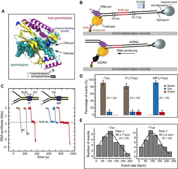
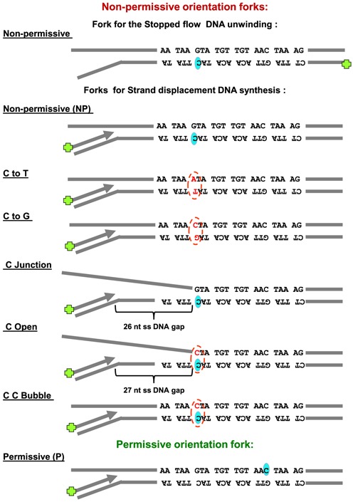
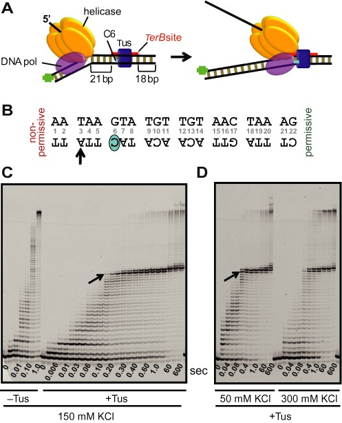
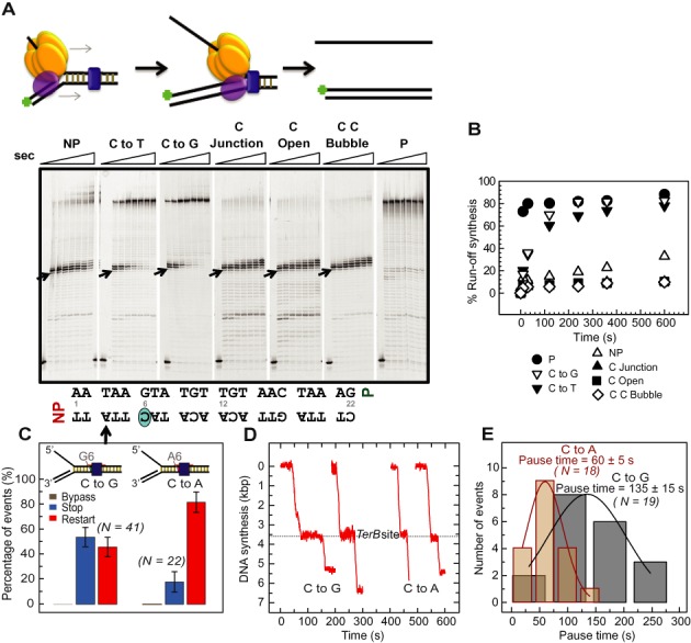
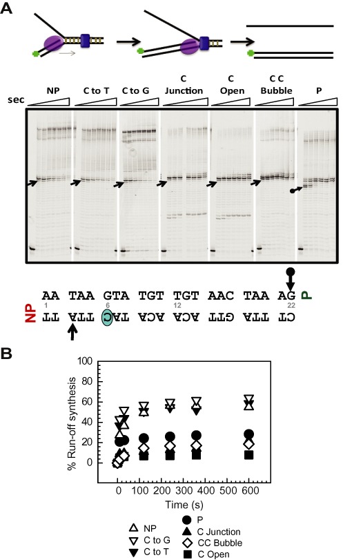
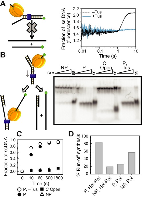
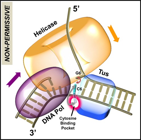
The Escherichia coli replication terminator protein (Tus) binds to Ter sequences to block replication forks approaching from one direction. Here, we used single molecule and transient state kinetics to study responses of the heterologous phage T7 replisome to the Tus-Ter complex. The T7 replisome was arrested at the non-permissive end of Tus-Ter in a manner that is explained by a composite mousetrap and dynamic clamp model. An unpaired C(6) that forms a lock by binding into the cytosine binding pocket of Tus was most effective in arresting the replisome and mutation of C(6) removed the barrier. Isolated helicase was also blocked at the non-permissive end, but unexpectedly the isolated polymerase was not, unless C(6) was unpaired. Instead, the polymerase was blocked at the permissive end. This indicates that the Tus-Ter mechanism is sensitive to the translocation polarity of the DNA motor. The polymerase tracking along the template strand traps the C(6) to prevent lock formation; the helicase tracking along the other strand traps the complementary G(6) to aid lock formation. Our results are consistent with the model where strand separation by the helicase unpairs the GC(6) base pair and triggers lock formation immediately before the polymerase can sequester the C(6) base.


Figures

Figure 1.
Figure 1.
Single-molecule DNA synthesis by T7 helicase–polymerase upon encountering the permissive or non-permissive face of the Tus–TerB complex. (A) Crystal structure (PDB code: 2I06) of the locked Tus–Ter complex shows the flipped C(6) base at the non-permissive face (5). (B) A schematic representation of the single-molecule tethered-bead experimental setup for observing DNA synthesis by the T7 helicase–polymerase. DNA synthesis converts the surface-tethered dsDNA to ssDNA, which at a regime force of 2.6 pN results in shortening of the DNA and displacement of the bead in the opposite direction to the flow. (C) Representative trajectories of DNA synthesis upon encountering Tus–TerB complexes. The fork primarily bypassed Tus–TerB complex on arrival at the permissive face (P, left), while it fully stopped at the non-permissive face (NP, right). Trajectories show permanent stoppage (in blue), unimpeded bypass (gray) or restart (red) at P or NP Tus–TerB. The location of the TerB site is indicated. Traces have been offset on the time axis for clarity. (D) The percentage of populations of replication forks that bypassed, transiently stopped or fully stopped at P TerB and NP TerB are shown. A control experiment in the absence of Tus is shown for NP TerB. Error bars correspond to the standard deviation of binomial distributions. (E) Rate of leading strand synthesis using forked-λ DNA in the absence (left) or presence of Tus (right). The rate distributions were fit (lines in black) with a Gaussian distribution. The uncertainties correspond to the standard error of the distribution. Leading-strand replication reactions were carried out in the presence or in the absence of Tus protein in buffer containing 50 mM potassium glutamate.
Figure 2.
Figure 2.
Schematic representation of the DNA fork substrates used in the bulk studies. TerB sequence orientation is shown pertaining to the DNA synthesis direction on primer. C(6) base (shaded oval shape) on Ter B sequence and any mutation of GC(6) is indicated (broken line oval shape).
Figure 3.
Figure 3.
Tus–TerB arrest of DNA synthesis by T7 helicase–DNA polymerase at single-nucleotide resolution. (A) Schematic of the experimental design to study replication arrest of T7 helicase-polymerase by Tus–TerB at single-nucleotide resolution using the chemical quenched flow assay. (B) The TerB sequence and the C(6) base. The TerB sequence numbering is followed throughout. (C) High resolution DNA sequencing gel shows progressive strand displacement DNA synthesis by the T7 helicase–polymerase on a fork DNA containing TerB in the non-permissive orientation. Arrows indicate the first arrest position band corresponding to the arrow on the TerB sequence in (B). These reactions were carried out in the quenched flow apparatus (QF) in the presence or in the absence of Tus protein at 150 mM KCl at 0.1 mM dVTPs and 1 mM dTTP. (D) Sequencing gel shows the QF reactions in the presence of Tus at 50 and 300 mM KCl, with all dNTPs at 1 mM. Each time point shown here is an independent reaction. Another QF experiment is also shown in Supplementary Figure S3 and extended time scale experiments are shown in Supplementary Figures S4, S6 and S7.
Figure 4.
Figure 4.
Strand displacement DNA synthesis by T7 helicase–polymerase on variant Tus–Ter fork DNAs. (A) T7 helicase and T7 DNA polymerase were preincubated with the preformed replication fork substrate and reacted with 1 mM dNTPs. DNA forks: non-permissive (NP), GC(6) to AT mutation (C to T), GC(6) to CG mutation (C to G), GC(6) at the junction (C Junction), C Open, C C Bubble and permissive (P). Reactions were quenched at 0, 10, 30, 120, 240, 360, 600, 1800 s and products were resolved in sequencing gels. Arrows indicates the first arrest position band corresponding to the arrow on the TerB sequence below. (B) Plot showing ‘% run-off synthesis’ against time, quantified from the gels in (A) as described in ‘Materials and Methods’ section. (C) Percentages of the populations of replication forks that bypassed (gray), transiently stopped (red) or fully stopped (blue) at Tus bound to TerB sites bearing the GC(6) to CG (C to G; left) or GC(6) to TA mutation (C to A; right). Error bars correspond to the standard deviation of binomial distributions. (D) Representative single molecule trajectories of the restart of DNA synthesis after transient stoppage at CG(6)-NP and TA(6)-NP TerB are shown. (E) The pause durations for events that restarted at CG(6)-NP or TA(6)-NP TerB were fit with Gaussian distributions (black and red lines, respectively). The repeat experiments carried out at 23°C are shown in Supplementary Figure S6–S9.
Figure 5.
Figure 5.
Strand displacement DNA synthesis by T7 DNA polymerase alone on various Tus–Ter fork DNAs. (A) T7 DNA polymerase was preincubated with the preformed replication fork substrate and reacted with 1 mM dNTPs. The DNA forks used here were as follows: non-permissive (NP), GC(6) to AT mutation (C to T), GC(6) to CG mutation (C to G), GC(6) at the junction (C Junction), C Open, C C Bubble and permissive (P). Reactions were quenched at 0, 10, 30, 120, 240, 360, 600, 1800 s and products resolved in sequencing gels. Arrows indicates the first arrest position band corresponding to the arrows on the TerB sequence below. (B) Plot showing ‘% run-off synthesis’ against time, quantified from the sequencing gels in (A). The duplicate sets of experiments are shown in Supplementary Figure S10 and also those conducted at 23°C in Supplementary Figure S9.
Figure 6.
Figure 6.
DNA unwinding by T7 helicase alone on various Tus–Ter fork DNAs. (A) Cartoon depiction of the stopped-flow fluorescence unwinding assay design (left). Fluorescence intensity traces represent the unwound ssDNA fraction as the result of DNA unwinding activity of T7 helicase on a non-permissive (NP) fork in the presence and absence of Tus (right), plotted against time. The plots are average of at least five reactions. (B) Radiometric gel unwinding assay showing helicase unwinding activity in the presence of Tus on non-permissive (NP), C Open, permissive (P) forks and on permissive (P) DNA forks in the absence of Tus. Reactions were quenched at 0, 10, 60, 600, 1800 s and then resolved in a non-denaturing PAGE gel. Lanes marked ‘ss’ represent single-stranded labeled DNA. (C) Plot showing unwound ssDNA fraction against time, quantified as described in ‘Materials and Methods’ section. (D) Comparison of run-off synthesis by helicase–DNA polymerase and DNA polymerase on fork DNAs with dsTerB in the non-permissive (NP) and permissive (P) orientations. The duplicate sets of experiments are shown in Supplementary Figure S11.
Figure 7.
Figure 7.
Model for replication fork arrest. The helicase ring and DNA polymerase are bound at the fork junction containing the Tus–Ter complex. After the Ter sequence is unwound up to the GC(6) base pair, the unpaired C(6) base can flip out and bind into the cytosine-specific pocket on Tus to form the C(6) Tus–Ter lock, or reanneal back with its complementary base G(6), or become base-paired through DNA synthesis. The ‘pin’ for strand separation occurs when C(6) on the template strand is already within three or fewer nucleotides of the polymerase active site. When the helicase-polymerase complex unwinds DNA, the helicase traps the G(6) base to prevent reannealing of GC(6) base pair, aiding formation of the locked complex. If GC(6) base pair is unwound by the strand displacement activity of the isolated polymerase, the C(6) is captured by the polymerase through DNA synthesis, which prevents the formation of the locked complex.

Similar articles

Cited by

References

    1. Hiasa H., Marians K.J. Tus prevents overreplication of oriC plasmid DNA. J. Biol. Chem. 1994;269:26959–26968. - PubMed
    1. Neylon C., Kralicek A.V., Hill T.M., Dixon N.E. Replication termination in Escherichia coli: structure and antihelicase activity of the Tus-Ter complex. Microbiol. Mol. Biol. Rev. 2005;69:501–526. - PMC - PubMed
    1. Gottlieb P.A., Wu S., Zhang X., Tecklenburg M., Kuempel P., Hill T.M. Equilibrium, kinetic, and footprinting studies of the Tus-Ter protein-DNA interaction. J. Biol. Chem. 1992;267:7434–7443. - PubMed
    1. Neylon C., Brown S.E., Kralicek A.V., Miles C.S., Love C.A., Dixon N.E. Interaction of the Escherichia coli replication terminator protein (Tus) with DNA: a model derived from DNA-binding studies of mutant proteins by surface plasmon resonance. Biochemistry. 2000;39:11989–11999. - PubMed
    1. Mulcair M.D., Schaeffer P.M., Oakley A.J., Cross H.F., Neylon C., Hill T.M., Dixon N.E. A molecular mousetrap determines polarity of termination of DNA replication in E. coli. Cell. 2006;125:1309–1319. - PubMed

Publication types