

ZnO Nanostructure-Based Intracellular Sensor
- PMID: 26007730
- PMCID: PMC4481971
- DOI: 10.3390/s150511787
ZnO Nanostructure-Based Intracellular Sensor
Abstract
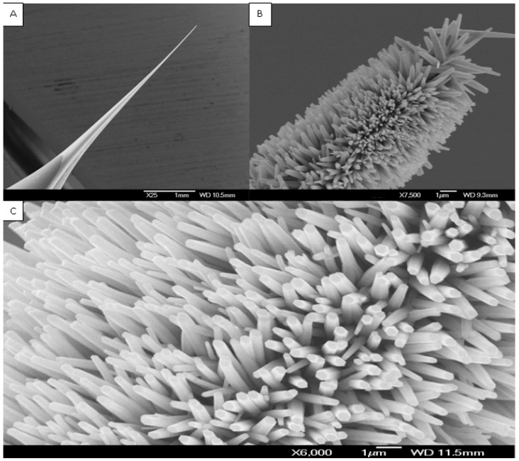
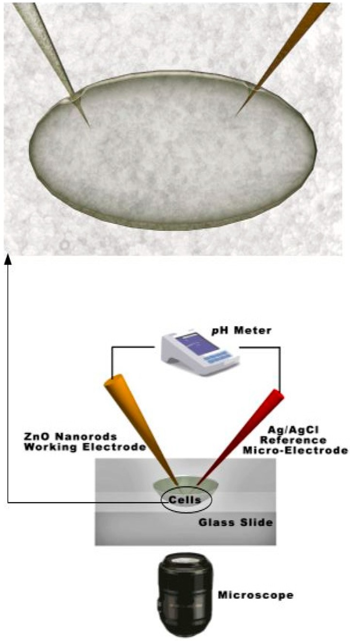
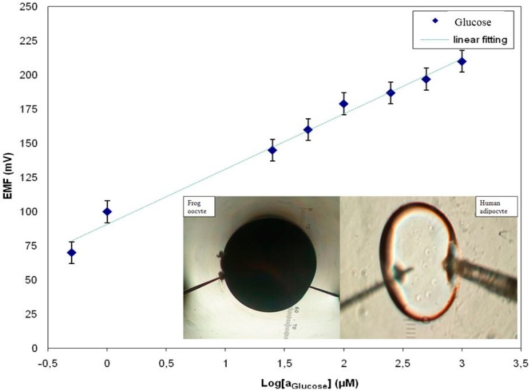
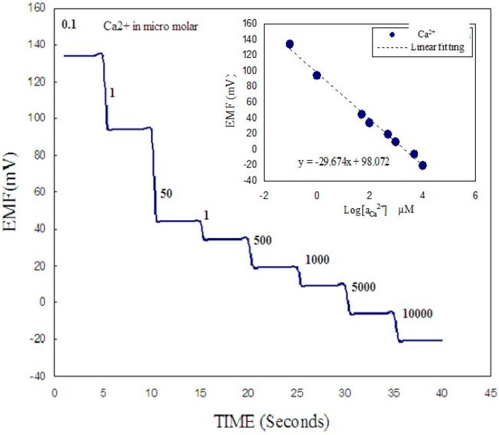
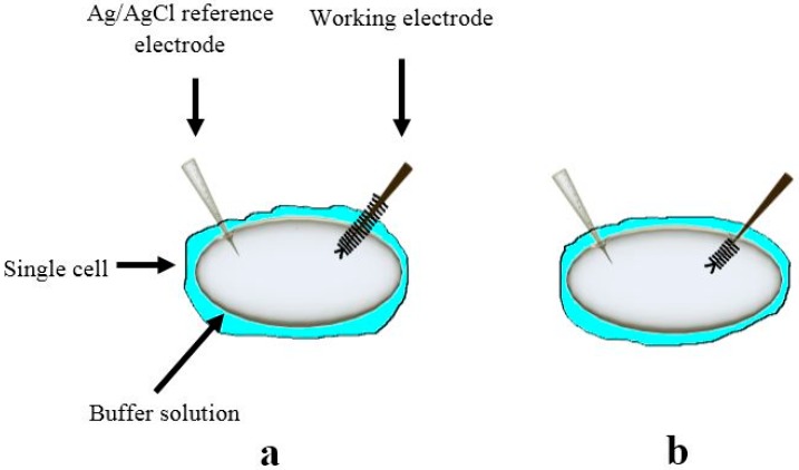
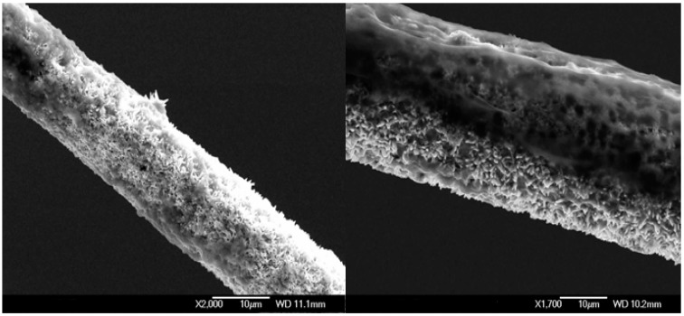
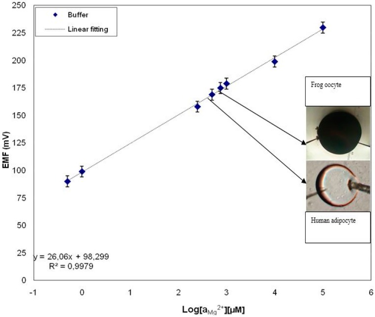
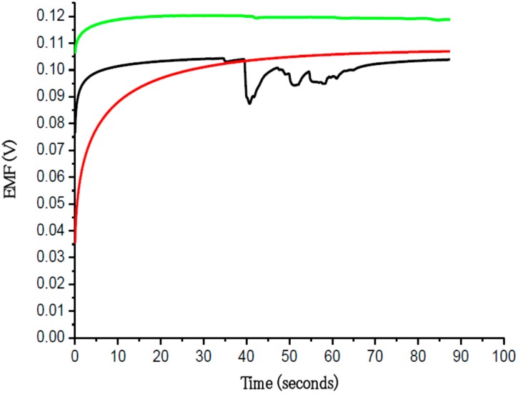
Recently ZnO has attracted much interest because of its usefulness for intracellular measurements of biochemical species by using its semiconducting, electrochemical, catalytic properties and for being biosafe and biocompatible. ZnO thus has a wide range of applications in optoelectronics, intracellular nanosensors, transducers, energy conversion and medical sciences. This review relates specifically to intracellular electrochemical (glucose and free metal ion) biosensors based on functionalized zinc oxide nanowires/nanorods. For intracellular measurements, the ZnO nanowires/nanorods were grown on the tip of a borosilicate glass capillary (0.7 µm in diameter) and functionalized with membranes or enzymes to produce intracellular selective metal ion or glucose sensors. Successful intracellular measurements were carried out using ZnO nanowires/nanorods grown on small tips for glucose and free metal ions using two types of cells, human fat cells and frog oocytes. The sensors in this study were used to detect real-time changes of metal ions and glucose across human fat cells and frog cells using changes in the electrochemical potential at the interface of the intracellular micro-environment. Such devices are helpful in explaining various intracellular processes involving ions and glucose.
Keywords: ZnO nanowire/nanorods, functionalization, intracellular measurement, glucose, metal ions, human fat cells, frog oocytes, electrochemical sensor.
Figures
Similar articles
-
Functionalized ZnO nanorod-based selective magnesium ion sensor for intracellular measurements.Biosens Bioelectron. 2010 Nov 15;26(3):1118-23. doi: 10.1016/j.bios.2010.08.017. Epub 2010 Aug 20. Biosens Bioelectron. 2010. PMID: 20846846
-
Functionalised ZnO-nanorod-based selective electrochemical sensor for intracellular glucose.Biosens Bioelectron. 2010 Jun 15;25(10):2205-11. doi: 10.1016/j.bios.2010.02.025. Epub 2010 Mar 3. Biosens Bioelectron. 2010. PMID: 20303253
-
Selective calcium ion detection with functionalized ZnO nanorods-extended gate MOSFET.Biosens Bioelectron. 2009 Jul 15;24(11):3379-82. doi: 10.1016/j.bios.2009.04.011. Epub 2009 Apr 16. Biosens Bioelectron. 2009. PMID: 19442511
-
ZnO-based nanostructured electrodes for electrochemical sensors and biosensors in biomedical applications.Biosens Bioelectron. 2019 Sep 15;141:111417. doi: 10.1016/j.bios.2019.111417. Epub 2019 Jun 8. Biosens Bioelectron. 2019. PMID: 31202187 Review.
-
Protein biosensors based on polymer nanowires, carbon nanotubes and zinc oxide nanorods.Sensors (Basel). 2011;11(5):5087-111. doi: 10.3390/s110505087. Epub 2011 May 9. Sensors (Basel). 2011. PMID: 22163892 Free PMC article. Review.
Cited by
-
Nanomaterial-Based Therapy for Wound Healing.Nanomaterials (Basel). 2022 Feb 12;12(4):618. doi: 10.3390/nano12040618. Nanomaterials (Basel). 2022. PMID: 35214947 Free PMC article. Review.
-
Recent Progress in Lab-On-a-Chip Systems for the Monitoring of Metabolites for Mammalian and Microbial Cell Research.Sensors (Basel). 2019 Nov 18;19(22):5027. doi: 10.3390/s19225027. Sensors (Basel). 2019. PMID: 31752167 Free PMC article. Review.
-
Single-Cell Endoscopy for Multifunctional Live-Cell Molecular Analysis.Biosensors (Basel). 2025 Apr 11;15(4):244. doi: 10.3390/bios15040244. Biosensors (Basel). 2025. PMID: 40277557 Free PMC article. Review.
-
Highly Sensitive Magnesium-Doped ZnO Nanorod pH Sensors Based on Electrolyte-Insulator-Semiconductor (EIS) Sensors.Sensors (Basel). 2021 Mar 17;21(6):2110. doi: 10.3390/s21062110. Sensors (Basel). 2021. PMID: 33802968 Free PMC article.
-
EGFET-Based Sensors for Bioanalytical Applications: A Review.Sensors (Basel). 2018 Nov 20;18(11):4042. doi: 10.3390/s18114042. Sensors (Basel). 2018. PMID: 30463318 Free PMC article. Review.
References
-
- Asif M.H., Willander M., Strålfors P., Danielsson B. In: Zinc Oxide Nanorods and Their Application to Intracellular Glucose Measurements. Le L.-A., Hunter R.J., Victor R., editors. Preedy Science Publishers, CRC; London, UK: 2012. pp. 120–140. Chapter 7: Nanotechnology and Nanomedicine in Diabetes.
-
- Arya A.K., Kumar L., Pukharia D., Tripathi K. Application of nanotechnology in diabetes. Dig. J. Nanomater. Biostruct. 2008;3:221–225.
-
- Asif M.H., Fulati A., Nur O., Willander M., Brännmark C., Strålfors P., Börjesson S.I., Elinder F. Functionalized zinc oxide nanorod with ionophore-membrane coating as an intracellular Ca2+ selective sensor. Appl. Phys. Lett. 2009;95:023703–023705. doi: 10.1063/1.3176441. - DOI
Publication types
MeSH terms
Substances
LinkOut - more resources
Full Text Sources
Other Literature Sources
