Review
. 2015 Jun;12(6):342-52.
doi: 10.1038/nrgastro.2015.74. Epub 2015 May 26.

Pathogenesis of biliary atresia: defining biology to understand clinical phenotypes

Affiliations
Review

Pathogenesis of biliary atresia: defining biology to understand clinical phenotypes

Akihiro Asai et al. Nat Rev Gastroenterol Hepatol. 2015 Jun.

Abstract

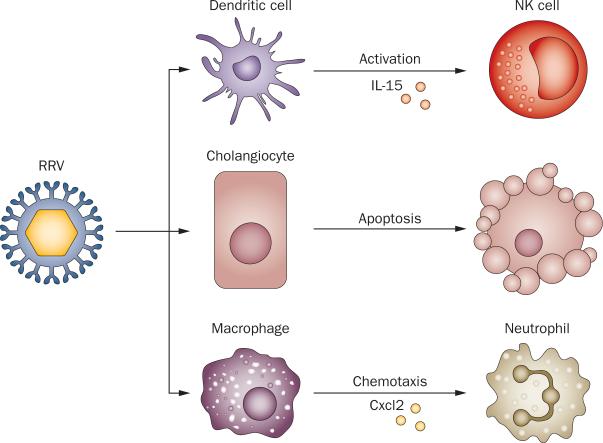
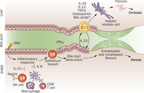
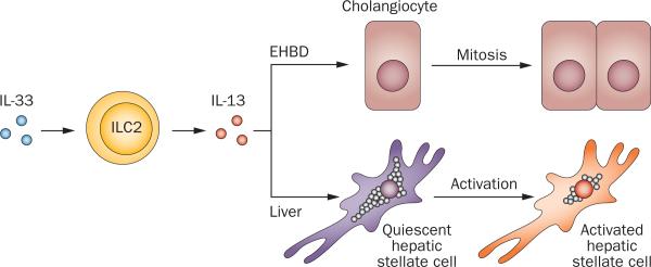
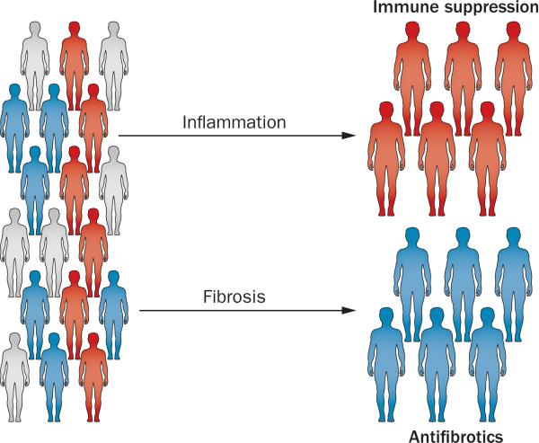
Biliary atresia is a severe cholangiopathy of early infancy that destroys extrahepatic bile ducts and disrupts bile flow. With a poorly defined disease pathogenesis, treatment consists of the surgical removal of duct remnants followed by hepatoportoenterostomy. Although this approach can improve the short-term outcome, the liver disease progresses to end-stage cirrhosis in most children. Further improvement in outcome will require a greater understanding of the mechanisms of biliary injury and fibrosis. Here, we review progress in the field, which has been fuelled by collaborative studies in larger patient cohorts and the development of cell culture and animal model systems to directly test hypotheses. Advances include the identification of phenotypic subgroups and stages of disease based on clinical, pathological and molecular features. Stronger evidence exists for viruses, toxins and gene sequence variations in the aetiology of biliary atresia, triggering a proinflammatory response that injures the duct epithelium and produces a rapidly progressive cholangiopathy. The immune response also activates the expression of type 2 cytokines that promote epithelial cell proliferation and extracellular matrix production by nonparenchymal cells. These advances provide insight into phenotype variability and might be relevant to the design of personalized trials to block progression of liver disease.


Figures

Figure 1
Figure 1
Cellular targets and molecular events after RRV infection. When administered into BALB/c mice in the first 2 postnatal days, RRV infects cholangiocytes, inducing apoptosis and necrosis. RRV also targets hepatic DCs, which activate NK cells via Il-15 and hepatic macrophages, which secrete Cxcl2 that attracts neutrophils. Abbreviations: Cxcl2, C-X-C motif chemokine 2; NK, natural killer cell; RRV, Rhesus rotavirus type A.
Figure 2
Figure 2
Biological processes and effectors of injury in biliary atresia. The release of IL-15 by DCs after RRV infection or toxin activates NK cells and CD8+ T cells. Targeting of cholangiocytes by infection or toxin activates inflammatory cells and the release of IL-15, perforin and granzymes, which injures the epithelium. IFN-γ expression is linked to an amplification of epithelial injury and duct obstruction by prominent infiltration of lymphocytes and myeloid cells. The release of IL-33 and IL-13 by ILC2 promotes epithelial repair in bile ducts, and is linked to fibrosis in the liver (and perhaps of extrahepatic ducts). Abbreviations: DC, dendritic cell; EHBD, extrahepatic bile duct; ILC2, innate lymphocyte cell type 2; M, macrophage; N, neutrophil; NK, natural killer cell; RRV, Rhesus rotavirus type A.
Figure 3
Figure 3
Epithelial and fibrogenic response induced by IL-33. The presence of IL-33 activates type 2 innate lymphoid cells (ILC2) to release IL-13, which induces cholangiocyte proliferation in the neighbouring epithelium of extrahepatic bile ducts. In the liver, the IL-33–ILC2–IL-13 circuit activates the profibrogenic phenotype of hepatic stellate cells. Abbreviation: EHBD, extrahepatic bile duct.
Figure 4
Figure 4
Designing clinical trials based on biological stages of liver disease. Histopathology and molecular methods to stage the liver disease might identify predominantly inflammation (red), fibrosis (blue) and mixed (grey) subgroups of patients at diagnosis. Matching of the disease stages with specific therapies in future trials could yield improved efficacy and prevent unnecessary exposure of patients to drug toxicity.

References

    1. Dehghani SM, Efazati N, Shahramian I, Haghighat M, Imanieh MH. Evaluation of cholestasis in Iranian infants less than three months of age. Gastroenterol. Hepatol. Bed Bench. 2015;8:42–48. - PMC - PubMed
    1. Hoerning A, et al. Diversity of disorders causing neonatal cholestasis—the experience of a tertiary pediatric center in Germany. Front. Pediatr. 2014;2:65. - PMC - PubMed
    1. Lee WS, Chai PF, Boey CM, Looi LM. Aetiology and outcome of neonatal cholestasis in Malaysia. Singapore Med. J. 2010;51:434–439. - PubMed
    1. Stormon MO, Dorney SF, Kamath KR, O’Loughlin EV, Gaskin KJ. The changing pattern of diagnosis of infantile cholestasis. J. Paediatr. Child Health. 2001;37:47–50. - PubMed
    1. Schreiber RA, Kleinman RE. Biliary atresia. J. Pediatr. Gastroenterol. Nutr. 2002;35(Suppl. 1):S11–S16. - PubMed

Publication types