. 2015 May 26:6:7215.
doi: 10.1038/ncomms8215.

Acetylation of Beclin 1 inhibits autophagosome maturation and promotes tumour growth

Affiliations

Acetylation of Beclin 1 inhibits autophagosome maturation and promotes tumour growth

Ting Sun et al. Nat Commun. .

Abstract

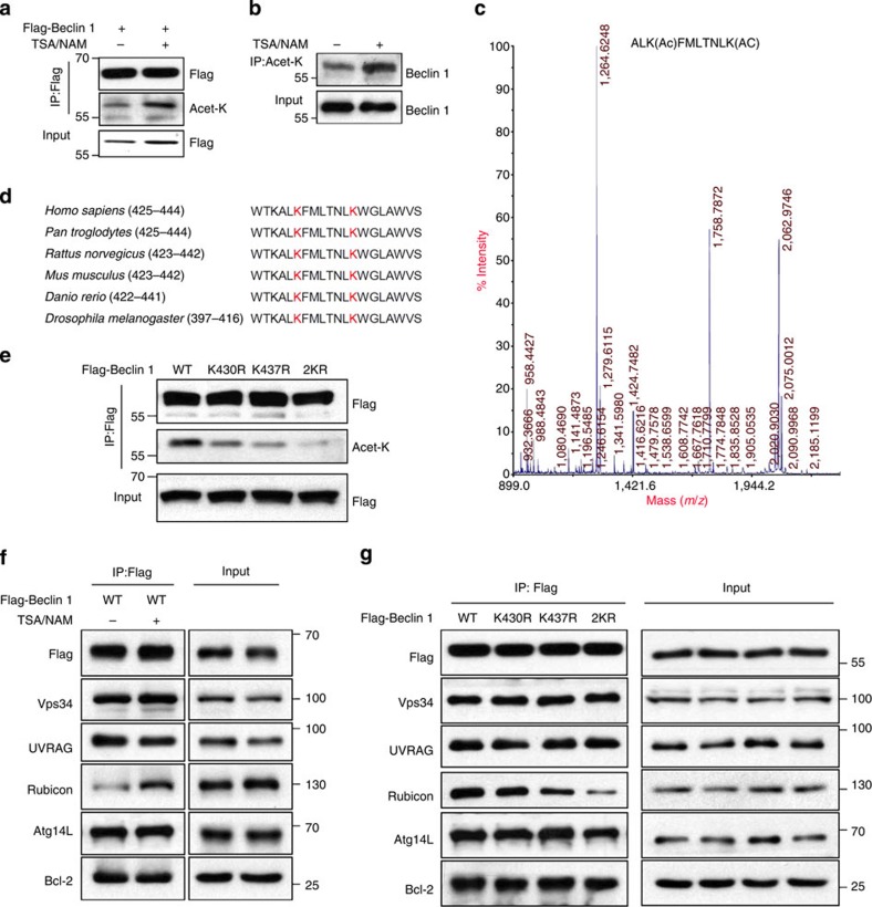
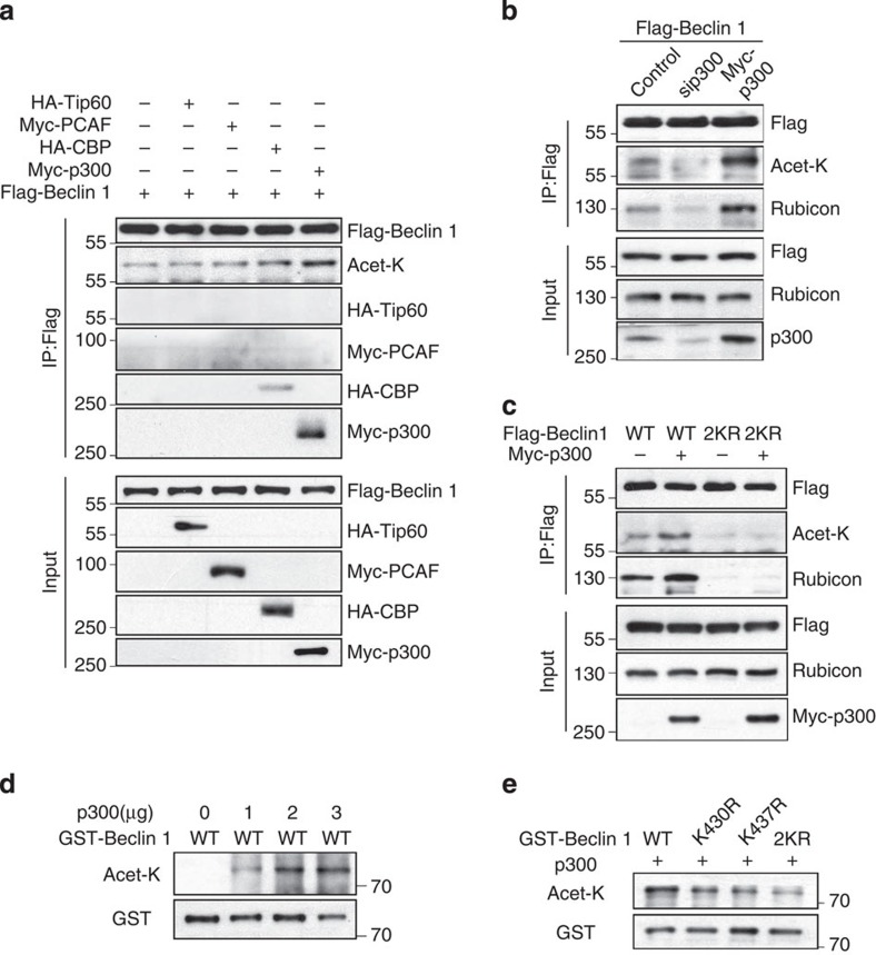
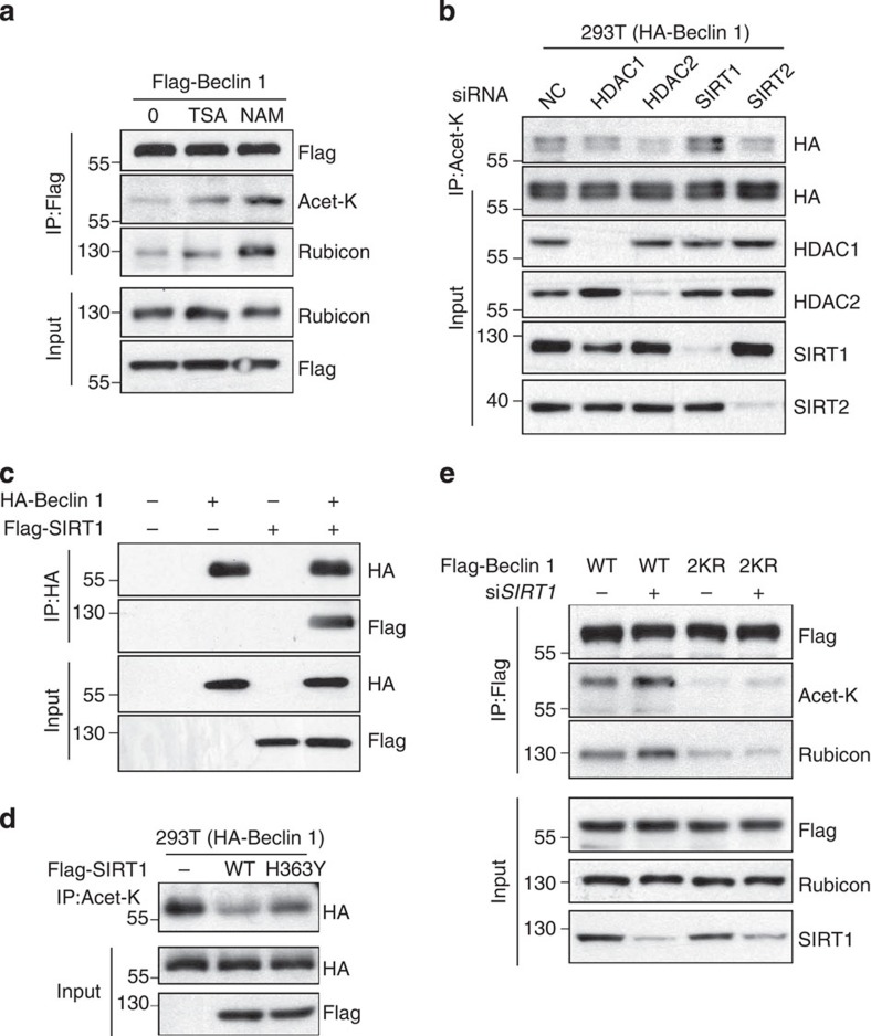
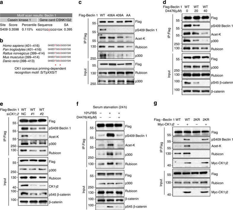
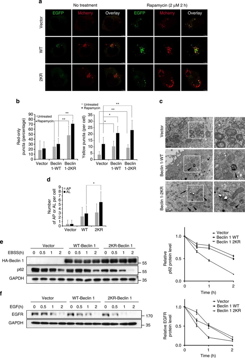
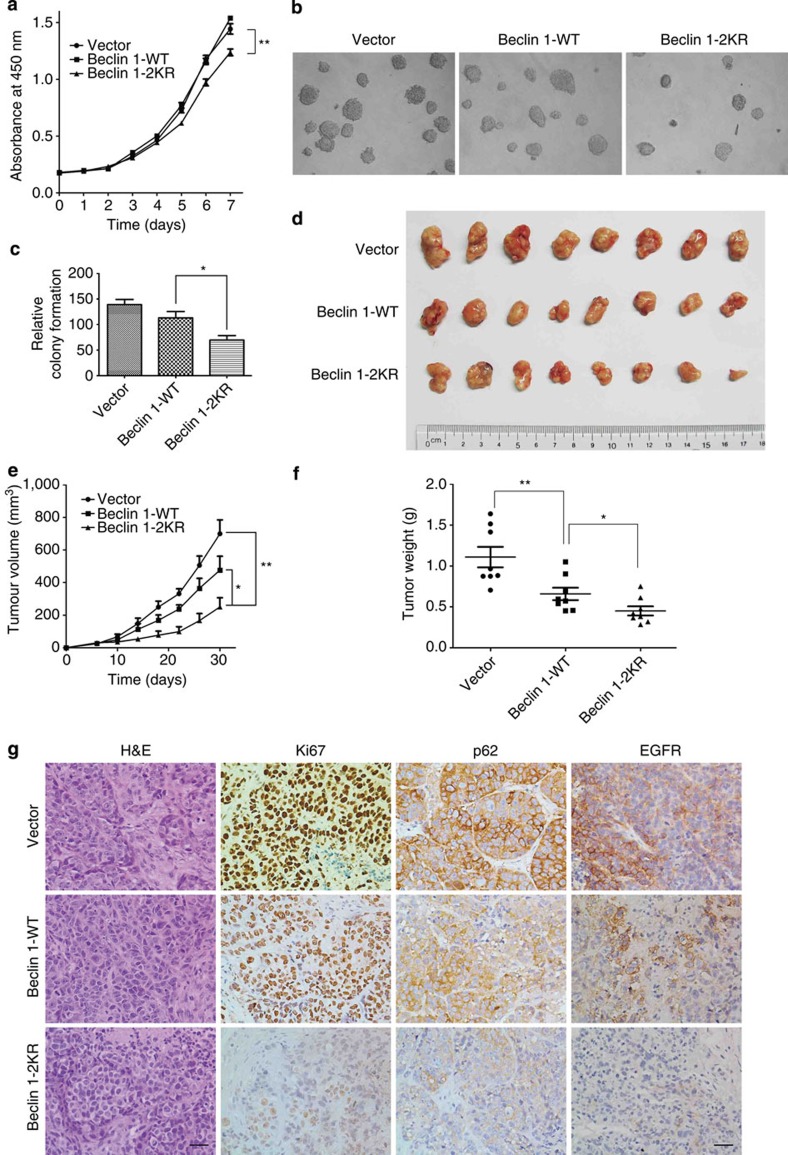
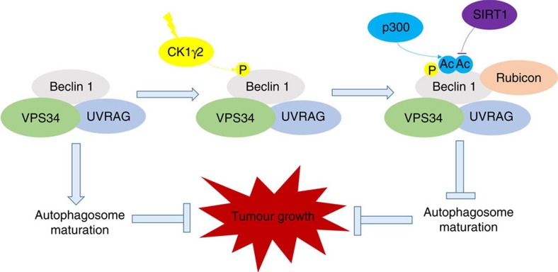
Beclin 1, a protein essential for autophagy, regulates autophagy by interacting with Vps34 and other cofactors to form the Beclin 1 complex. Modifications of Beclin 1 may lead to the induction, inhibition or fine-tuning of the autophagic response under a variety of conditions. Here we show that Beclin 1 is acetylated by p300 and deacetylated by SIRT1 at lysine residues 430 and 437. In addition, the phosphorylation of Beclin 1 at S409 by CK1 is required for the subsequent p300 binding and Beclin 1 acetylation. Beclin 1 acetylation inhibits autophagosome maturation and endocytic trafficking by promoting the recruitment of Rubicon. In tumour xenografts, the expression of 2KR mutant Beclin 1 (substitution of K430 and K437 to arginines) leads to enhanced autophagosome maturation and tumour growth suppression. Therefore, our study identifies an acetylation-dependent regulatory mechanism governing Beclin 1 function in autophagosome maturation and tumour growth.


Figures

Figure 1
Figure 1. Beclin 1 is acetylated at lysines 430 and 437.
(a) Exogenous Beclin 1 is acetylated. Acetylation of immunoprecipitated Flag-tagged Beclin 1 from HEK293T cells treated with or without HDAC inhibitors TSA (1 μM) and NAM (5 mM) simultaneously for 6 h. (b) Endogenous Beclin 1 is acetylated. Acetylated proteins were immunoprecipitated with the antibody to acetylated lysine from HEK293T cells after TSA and NAM treatments as indicated. Acetylation of endogenous Beclin 1 protein was analysed with western bolt analysis. (c) Identification of Beclin 1 K430 and K437 acetylation using mass spectrometry analysis. Flag-tagged Beclin 1 was transfected into HEK293T cells. At 24 h post transfection, TSA (1 μM) and NAM (5 mM) were added for another 6 h. Beclin 1 was purified by immunoprecipitation with an anti-Flag antibody and then analysed using mass spectrometry. (d) Alignment of the protein sequences of Beclin 1 homologues in various species. The red indicates the identified acetylated lysine residues of Beclin 1. (e) Mutations of K430 and K437 decrease Beclin 1 acetylation. Acetylation of ectopically expressed WT, K430R, K437R and Beclin 1–2KR was analysed. (f) TSA and NAM increase the binding of Beclin 1 to Rubicon. Immunoprecipitation of indicated Beclin 1-binding partners with ectopically expressed Flag-Beclin 1 in HEK293T cells treated with TSA and NAM. (g) Mutations of K430 and K437 decrease the binding of Beclin 1 and Rubicon. Immunoprecipitation of indicated proteins with Beclin 1 in HEK293T cells transfected with indicated Beclin 1 constructs.
Figure 2
Figure 2. p300 is the acetyltransferase of Beclin 1.
(a) Overexpression of p300, but not other acetyltransferases (HATs), could significantly bind with Beclin 1 and increase Beclin 1 acetylation. HA-tagged CBP or Tip60 or Myc-tagged p300 or p300/CBP-associated factor (PCAF) was co-transfected individually with Flag-Beclin 1 into HEK293T cells. (b) Knockdown or overexpression of p300 alters Beclin 1 acetylation and Beclin 1–Rubicon interaction. Flag-Beclin 1 was co-transfected with sip300, Myc-p300 or vector into HEK293T cells, Beclin 1 acetylation and the interaction between Beclin 1 and Rubicon was determined with IP and western blot analyses. (c) Deacetylation mimic (2KR) counteracts p300 acetylation of Beclin 1 and diminishes the binding of Beclin 1 with Rubicon. Flag-tagged Beclin 1 (WT, 2KR) was co-transfected with Myc-p300 or vector into HEK293T cells. (d) Beclin 1 is acetylated by p300 in vitro. Different doses of recombinant human p300 were incubated with bacterially expressed GST–Beclin 1 in the presence of 100 μM AcCoA for 1 h at 30 °C. (e) Sequential mutation of two acetylable lysine residues (K430 and K437) to arginine progressively diminished the acetylation of GST–Beclin 1 by p300 in vitro.
Figure 3
Figure 3. SIRT1 decreases Beclin 1 acetylation and the interaction of Beclin 1 and Rubicon.
(a) NAM, not TSA, increases Beclin 1 acetylation. Flag-tagged Beclin 1 was transfected into HEK293T cells with or without sirtuin deacetylase inhibitor NAM and HDAC inhibitor TSA. Acetylation of Beclin 1 was measured with IP western blot analyses. (b) SIRT1 knockdown increases Beclin 1 acetylation. HEK293T cells stably expressing HA-Beclin 1 were transfected with siRNA targeting HDAC1, HDAC2, SIRT1 and SIRT2 or negative control. Beclin 1 acetylation was measured by immunoprecipitation using an anti-acetylated lysine antibody. (c) Association of Beclin 1 with SIRT1. HA-tagged Beclin 1 and Flag-tagged p300 were transfected into HEK293T cells individually or together. The interaction between Beclin 1 and SIRT1 was determined with IP and western blot analyses. (d) Catalytic activity of SIRT1 is required for deacetylation of Beclin 1. HEK293T cells stably expressing HA-Beclin 1 were transfected into Flag-tagged SIRT1 WT or catalytically inactive mutant H363Y. Beclin 1 acetylation was measured by immunoprecipitation using an anti-acetylated lysine antibody. (e) Beclin 1–2KR-mutant counteracts siSIRT1 acetylation of Beclin 1 and diminishes the Beclin 1–Rubicon interaction. Flag-tagged Beclin 1 (WT, 2KR) was co-transfected with siSIRT1 or negative control into HEK293T cells.
Figure 4
Figure 4. CK1-mediated phosphorylation of Beclin 1 is essential for Beclin 1 acetylation.
(a) Motif scan result of Beclin 1 by Scansite. Choosing the stringency level is high. (b) Cluster alignment of CK1 consensus phosphorylation motif on Beclin 1 proteins. Arrowheads point to T406 and S409 of human Beclin 1. (c) Mutations of T406 and S409 decrease Beclin 1 phosphorylation and acetylation. HEK293T cells were transfected with indicated Beclin 1 constructs. Beclin 1 phosphorylation, acetylation and the Beclin 1–Rubicon interaction were detected by IP as indicated. (d) Inhibition of CK1 with D4476 decreases Beclin 1 acetylation. HEK293T cells transfected with Flag-Beclin 1 were treated with indicated concentration of D4476 for 4 h. Beclin 1 acetylation and the interaction between Beclin 1 and p300 or Rubicon was determined with IP and western blot analyses. (e) CK1γ2 knockdown decreases the binding of p300 to Beclin 1 and Beclin 1 acetylation. Flag-Beclin 1 was co-transfected with siRNA into HEK293T cells. Beclin 1 acetylation and the interaction between Beclin 1 and p300 or Rubicon was determined with IP and western blot analyses. (f) Serum restoration increases Beclin 1 acetylation by activating CK1. HEK293T cells transfected with Flag-Beclin 1 were serum-starved for 24 h, and then serum was restored with or without D4476 pretreatment (40 μM, 2 h). (g) Overexpression of CK1γ2 increases WT Beclin 1 acetylation but not 2KR mutant. Flag-tagged Beclin 1 (WT, 2KR) was co-transfected with Myc-CK1γ2 or vector into HEK293T cells.
Figure 5
Figure 5. Autophagosome maturation and endocytic trafficking in Beclin 1–2KR-expressing cells.
(a) Beclin 1–2KR mutant promotes the maturation of autophagosomes. 293T cells stably expressing Vector control, Beclin 1 WT or 2KR were transfected with mCherry-EGFP-LC3 in the absence or presence of rapamycin (2 μM). Scale bars, 5 μm. (b) Quantitation of mCherry-LC3-only puncta and the mCherry-EGFP overlay puncta in 293T cell lines treated as in a. Bars represent mean±s.d. of 50 cells; five independent experiments, *P<0.05, **P<0.01 (Student's t-test). (c) Electron microscopy analysis of autophagosomes and autolysosomes. MCF7 cells stably expressing Beclin 1 (WT or 2KR) or Vector grown in normal medium was subjected to electron microscopy analysis. The arrows indicate autophagosomes (AP) and the arrowheads indicate autolysosomes (AL). Scale bar, 0.5 μm. (d) Quantitation of AP and AL in MCF7 cell lines as in c. Bar are mean ±s.d. of 20 cells; three independent experiments, *P<0.05 (Student's t-test). (e) The effect of Beclin 1–2KR mutant on p62 turnover. MCF7 cells stably expressing vector control, Beclin 1 WT or 2KR were treated with EBSS for the indicated times. The band intensity was measured in three independent experiments and the mean±s.d. are shown (down panel). (f) EGFR degradation. MCF7 cells stably expressing vector control, Beclin 1 WT or 2KR were serum-starved for 24 h, and then treated with EGF (200 ng ml−1) for the indicated times. The band intensity was measured in three independent experiments and the mean±s.d. are shown (down panel).
Figure 6
Figure 6. Acetylation of Beclin 1 promotes tumour cell proliferation and tumorigenesis in vivo.
(a) Beclin 1–2KR mutant inhibits cell proliferation. MCF7 cells stably expressing vector control, Beclin 1 WT or 2KR were seeded at the same number in each well. Then made CCK-8 assay every 24 h. Results are mean±s.d. for experiments performed in triplicate, **P<0.01 (Student's t-test). (b) MCF7 cells stably expressing vector control, Beclin 1 WT or 2KR were used for colony formation assays. Cells (0.5 × 10⁴) were grown in soft agar, and the colonies were monitored after 2 weeks. Representative pictures of the colonies are shown. Results from three independent experiments are presented as means±s.d., *P<0.05 (Student's t-test). (d) Beclin 1–2KR mutant inhibits xenograft tumour growth. MCF7 cell lines (6 × 10⁶ cells) with vector control, Beclin 1 WT or 2KR were injected subcutaneously into the right flank of nude mice. At 38 days after injection, tumours were extracted and photographed. (e) Tumour growth curves in nude mice. Tumour diameters were measured at the indicated time points, and tumour volumes were calculated. Results are mean volume±s.e.m. for 10 mice per group per time point. *P<0.05 (Student's t-test). (f) Tumour weights from experiment in c on autopsy on day 38. *P<0.05 (Student's t-test). (g) Beclin 1–2KR expression inhibits tumour cell proliferation in vivo. Tumour sections from xenografts were prepared for immunohistochemistry analysis. Representative haematoxylin and eosin staining (H&E) and Ki-67, p62, EGFR immunostaining images of indicated MCF7 xenograft tumour genotype. Scale bars, 20 μm.
Figure 7
Figure 7. A working model of Beclin 1 acetylation on autophagy and tumour growth.

References

    1. Mizushima N. & Komatsu M. Autophagy: renovation of cells and tissues. Cell 147, 728–741 (2011). - PubMed
    1. Levine B. & Klionsky D. J. Development by self-digestion: molecular mechanisms and biological functions of autophagy. Dev. Cell 6, 463–477 (2004). - PubMed
    1. Levine B. & Kroemer G. Autophagy in the pathogenesis of disease. Cell 132, 27–42 (2008). - PMC - PubMed
    1. Mizushima N., Levine B., Cuervo A. M. & Klionsky D. J. Autophagy fights disease through cellular self-digestion. Nature 451, 1069–1075 (2008). - PMC - PubMed
    1. Shintani T. & Klionsky D. J. Autophagy in health and disease: a double-edged sword. Science 306, 990–995 (2004). - PMC - PubMed

Publication types

MeSH terms