Review
. 2015 May 25;209(4):493-506.
doi: 10.1083/jcb.201412147.

Brain barriers: Crosstalk between complex tight junctions and adherens junctions

Affiliations
Review

Brain barriers: Crosstalk between complex tight junctions and adherens junctions

Silvia Tietz et al. J Cell Biol. .

Abstract

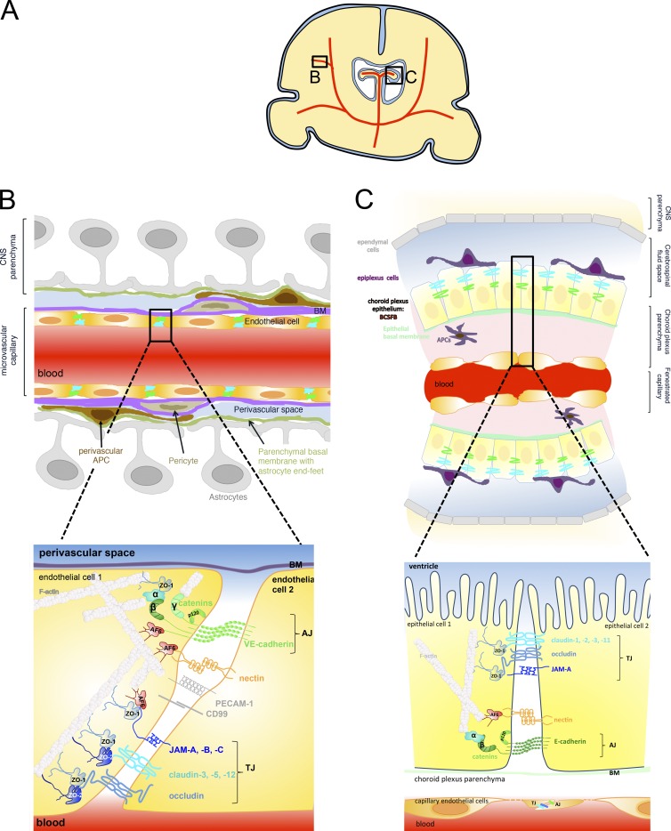
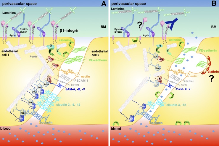
Unique intercellular junctional complexes between the central nervous system (CNS) microvascular endothelial cells and the choroid plexus epithelial cells form the endothelial blood-brain barrier (BBB) and the epithelial blood-cerebrospinal fluid barrier (BCSFB), respectively. These barriers inhibit paracellular diffusion, thereby protecting the CNS from fluctuations in the blood. Studies of brain barrier integrity during development, normal physiology, and disease have focused on BBB and BCSFB tight junctions but not the corresponding endothelial and epithelial adherens junctions. The crosstalk between adherens junctions and tight junctions in maintaining barrier integrity is an understudied area that may represent a promising target for influencing brain barrier function.


Figures

Figure 1.
Figure 1.
Intercellular junctions between CNS microvascular endothelial cells forming the BBB and between choroid plexus epithelial cells forming the BCSFB. (A) Schematic of the localization of the brain barriers in parenchymal microvessels (B) and the choroid plexus in the ventricles (C) in a coronal brain section. (B) Localization (top) and molecular composition (bottom) of endothelial BBB AJs and TJs. AJs at the BBB are established by the homophilic interaction of cis dimers of transmembrane VE-cadherin in between adjacent endothelial cells. The cytoplasmic tail of VE-cadherin binds the armadillo family proteins p120- and β-catenin, which, via interaction with α-catenin and afadin (AF-6), mediate the link to the actin cytoskeleton. Nectins are transmembrane proteins belonging to the immunoglobulin (Ig) superfamily that form homodimers in cis and contribute to AJ formation by adhering to nectins on the adjacent cell and binding AF-6 via their cytoplasmic tail, which connects nectin with the actin cytoskeleton. VE-cadherin can also bind to plakoglobin (γ-catenin) instead of to β-catenin and thus connect endothelial AJs to the intermediate filaments in addition to the actin cytoskeleton. Although drawn separately in this figure, TJ strands that encircle the entire circumference of the CNS endothelial cells are intermingled with AJs. The transmembrane TJ proteins found to be localized in CNS endothelium are occludin, claudin-3, claudin-5, and claudin-12 as well as the junctional adhesion molecules (JAM) A, B, and C. Both claudins and JAMs mediate homophilic and heterophilic cis and trans interactions within their family, thus sealing the paracellular cleft between the adjacent endothelial cells. With the exception of claudin-12, the transmembrane TJ proteins carry a PDZ-binding motif in their carboxy terminus, through which they bind the scaffolding proteins ZO-1 and ZO-2 and, in the case of JAMs, also AF-6. These scaffolding proteins mediate the link to the actin cytoskeleton. ZO-1 has also been shown to bind to α-catenin and F-actin and thus regulate AJs. Additional molecules found to be present in BBB cell-to-cell contacts are the Ig supergene family members ESAM, PECAM-1, and CD99. Linkage of TJ proteins to cell polarity complexes are omitted from this figure for reasons of simplicity. (C) Localization (top) and molecular composition (bottom) of the epithelial BCSFB established by choroid plexus epithelial cells. The microvessels within the highly vascularized choroid plexus are fenestrated capillaries that allow the free diffusion of water-soluble molecules across the vascular wall. Nevertheless, these vascular endothelial cells form AJs and TJs resembling those of peripheral vascular beds and are thus not drawn in detail here. AJs of the BCSFB are formed by the homophilic interaction of cis homodimers of the transmembrane protein E-cadherin. Via its cytoplasmic tail, E-cadherin binds the armadillo family members p120- and β-catenin, which, in turn bind α-catenin, linking E-cadherin to the epithelial actin cytoskeleton. Oriented apically relative to the AJs, the TJ strands of the BCSFB run in parallel around the entire circumference of the choroid plexus epithelial cells, with characteristic gaps that have been suggested to be important in the context of the transport functions of the choroid plexus. The transmembrane proteins that have been characterized as localized to the BCSFB TJs include occludin, claudin-1, claudin-2, claudin-3, and claudin-11 as well as JAM-A and JAM-C. Via their carboxy terminus, which harbors a PDZ domain binding motif, these proteins bind the scaffolding proteins ZO-1, ZO-2, and probably ZO-3, which link to the actin cytoskeleton. BM, basement membrane.
Figure 2.
Figure 2.
Loss of endothelial–matrix interactions in neuroinflammation affects junctional integrity of the BBB. (A) Under physiological conditions, BBB endothelial cells are anchored via β1-integrins to the ECM proteins laminin, collagen, and perlecan in the endothelial basement membrane. β1-integrin–mediated adhesive contacts contribute by not-yet-defined downstream signaling events to junctional stabilization. (B) After ischemic stroke, edema formation and BBB breakdown is associated with loss of vascular β1-integrin, probably by shedding or degradation (indicated by a question mark). Genetic or functional inactivation of β1-integrin at the BBB induces BBB leakiness by reducing the association of p120 and β-catenin with VE-cadherin. This leads to increased internalization of VE-cadherin via clathrin-coated pits and ultimately loss of junctional claudin-5. BM, basement membrane.

Similar articles

Cited by

References

    1. Alvarez J.I., Dodelet-Devillers A., Kebir H., Ifergan I., Fabre P.J., Terouz S., Sabbagh M., Wosik K., Bourbonnière L., Bernard M., et al. . 2011. The Hedgehog pathway promotes blood-brain barrier integrity and CNS immune quiescence. Science. 334:1727–1731. 10.1126/science.1206936 - DOI - PubMed
    1. Alvarez J.I., Saint-Laurent O., Godschalk A., Terouz S., Briels C., Larouche S., Bourbonnière L., Larochelle C., and Prat A.. 2015. Focal disturbances in the blood-brain barrier are associated with formation of neuroinflammatory lesions. Neurobiol. Dis. 74:14–24. 10.1016/j.nbd.2014.09.016 - DOI - PubMed
    1. Argaw A.T., Gurfein B.T., Zhang Y., Zameer A., and John G.R.. 2009. VEGF-mediated disruption of endothelial CLN-5 promotes blood-brain barrier breakdown. Proc. Natl. Acad. Sci. USA. 106:1977–1982. 10.1073/pnas.0808698106 - DOI - PMC - PubMed
    1. Argaw A.T., Asp L., Zhang J., Navrazhina K., Pham T., Mariani J.N., Mahase S., Dutta D.J., Seto J., Kramer E.G., et al. . 2012. Astrocyte-derived VEGF-A drives blood-brain barrier disruption in CNS inflammatory disease. J. Clin. Invest. 122:2454–2468. 10.1172/JCI60842 - DOI - PMC - PubMed
    1. Armulik A., Genové G., Mäe M., Nisancioglu M.H., Wallgard E., Niaudet C., He L., Norlin J., Lindblom P., Strittmatter K., et al. . 2010. Pericytes regulate the blood-brain barrier. Nature. 468:557–561. 10.1038/nature09522 - DOI - PubMed

Publication types