

Retinal Vascular Changes are a Marker for Cerebral Vascular Diseases
- PMID: 26008809
- PMCID: PMC4743651
- DOI: 10.1007/s11910-015-0561-1
Retinal Vascular Changes are a Marker for Cerebral Vascular Diseases
Abstract
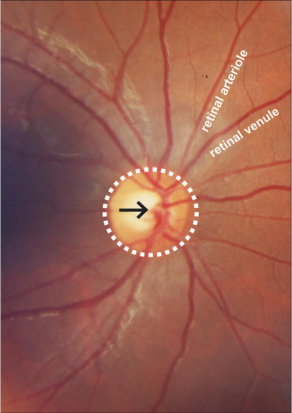
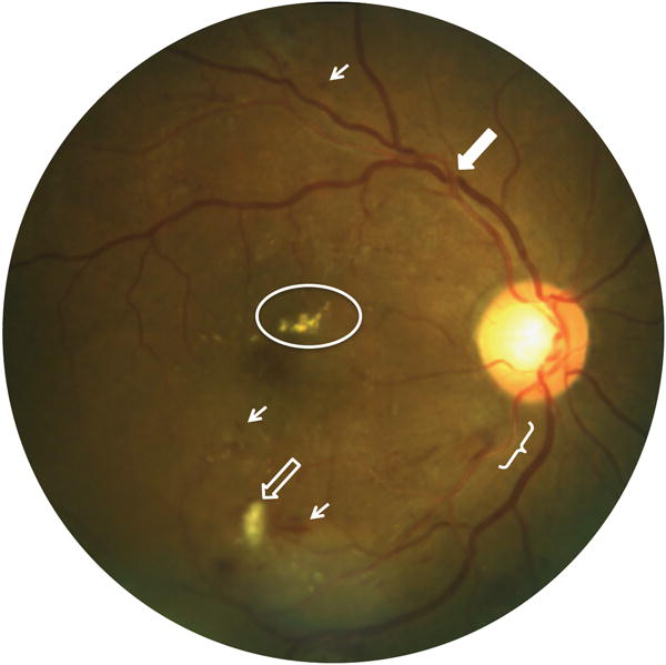
The retinal circulation is a potential marker of cerebral vascular disease because it shares origin and drainage with the intracranial circulation and because it can be directly visualized using ophthalmoscopy. Cross-sectional and cohort studies have demonstrated associations between chronic retinal and cerebral vascular disease, acute retinal and cerebral vascular disease, and chronic retinal vascular disease and acute cerebral vascular disease. In particular, certain qualitative features of retinopathy, retinal artery occlusion, and increased retinal vein caliber are associated with concurrent and future cerebrovascular events. These associations persist after accounting for confounding variables known to be disease-causing in both circulations, which supports the potential use of retinal vasculature findings to stratify individuals with regards to cerebral vascular disease risk.
Figures
References
-
- Delaey C, Van De Voorde J. Regulatory mechanisms in the retinal and choroidal circulation. Ophthalmic research. 2000;32(6):249–56. - PubMed
-
- Kwa VI, van der Sande JJ, Stam J, Tijmes N, Vrooland JL. Retinal arterial changes correlate with cerebral small-vessel disease. Neurology. 2002;59(10):1536–40. - PubMed
-
- Kalitzeos AA, Lip GY, Heitmar R. Retinal vessel tortuosity measures and their applications. Experimental eye research. 2013;106:40–6. - PubMed
-
- Cheung CY, Tay WT, Ikram MK, Ong YT, De Silva DA, Chow KY, et al. Retinal microvascular changes and risk of stroke: the Singapore Malay Eye Study. Stroke; a journal of cerebral circulation. 2013;44(9):2402–8. - PubMed
Publication types
MeSH terms
Grants and funding
LinkOut - more resources
Full Text Sources
Other Literature Sources
Medical
