. 2015 Jun 1;212(6):927-38.
doi: 10.1084/jem.20142384. Epub 2015 May 25.

Aberrant actin depolymerization triggers the pyrin inflammasome and autoinflammatory disease that is dependent on IL-18, not IL-1β

Affiliations

Aberrant actin depolymerization triggers the pyrin inflammasome and autoinflammatory disease that is dependent on IL-18, not IL-1β

Man Lyang Kim et al. J Exp Med. .

Abstract

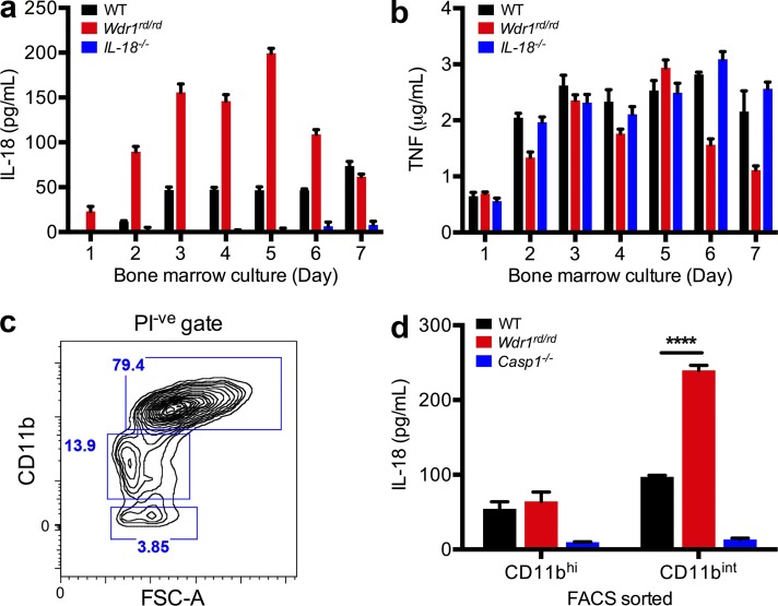
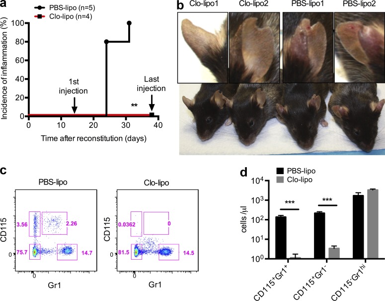
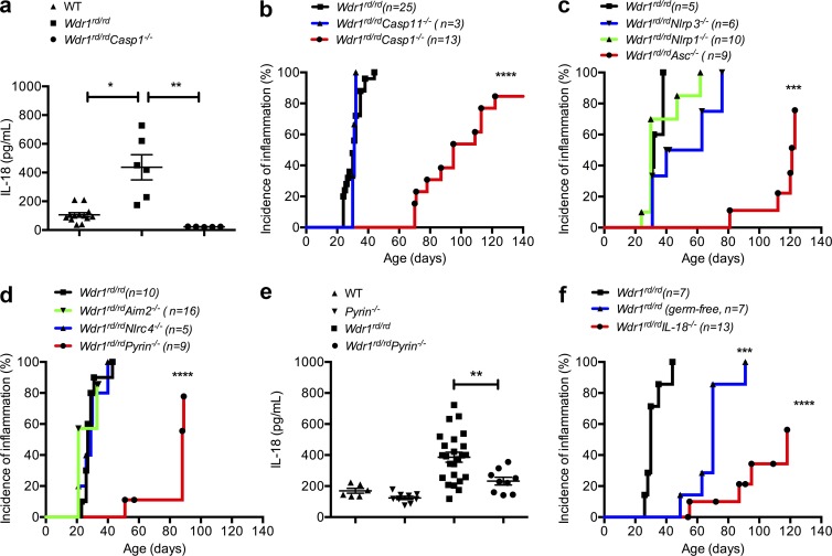
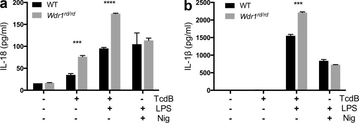
Gain-of-function mutations that activate the innate immune system can cause systemic autoinflammatory diseases associated with increased IL-1β production. This cytokine is activated identically to IL-18 by an intracellular protein complex known as the inflammasome; however, IL-18 has not yet been specifically implicated in the pathogenesis of hereditary autoinflammatory disorders. We have now identified an autoinflammatory disease in mice driven by IL-18, but not IL-1β, resulting from an inactivating mutation of the actin-depolymerizing cofactor Wdr1. This perturbation of actin polymerization leads to systemic autoinflammation that is reduced when IL-18 is deleted but not when IL-1 signaling is removed. Remarkably, inflammasome activation in mature macrophages is unaltered, but IL-18 production from monocytes is greatly exaggerated, and depletion of monocytes in vivo prevents the disease. Small-molecule inhibition of actin polymerization can remove potential danger signals from the system and prevents monocyte IL-18 production. Finally, we show that the inflammasome sensor of actin dynamics in this system requires caspase-1, apoptosis-associated speck-like protein containing a caspase recruitment domain, and the innate immune receptor pyrin. Previously, perturbation of actin polymerization by pathogens was shown to activate the pyrin inflammasome, so our data now extend this guard hypothesis to host-regulated actin-dependent processes and autoinflammatory disease.


Figures

Figure 1.
Figure 1.
Autoinflammatory disease in Wdr1rd/rd mice is IL-1 independent, but IL-18 dependent. (a and b) Serum cytokines/chemokines from wild-type (Wdr1+/+) and Wdr1rd/rd mice were analyzed by Bioplex (a) or for IL-1β and IL-18 by ELISA (b). n = 5–6. *, P < 0.05; **, P < 0.01; ***, P < 0.001 by unpaired t test, corrected for multiple comparisons. (c) Incidence of inflammation in tail or ears for Wdr1rd/rd, Wdr1rd/rdIL-1R−/−, or Wdr1rd/rdIL-18−/− mice. ****, P < 0.0001 by Gehan-Breslow-Wilcoxon test. (d) Incidence of inflammation after reconstitution of lethally irradiated mice with BM of the indicated genotypes (donor BM→irradiated host). ****, P < 0.0001 by Gehan-Breslow-Wilcoxon test. (e) Serum IL-18 from the mice shown in panel d was analyzed by ELISA. n = 4 or 6. Error bars represent means ± SEM. *, P < 0.05 by one-tailed unpaired t test. Except for disease incidence, all data are representative of two to four independent experiments.
Figure 2.
Figure 2.
LPS-induced IL-18 secretion from Wdr1rd/rd monocytes, not macrophages. (a and b) BM from WT, Wdr1rd/rd, or IL-18−/− mice was cultured in L929 cell-conditioned medium for 1–7 d. 105 cells from day 1–7 cultures were incubated with 1 µg/ml LPS. 48 h after stimulation, culture supernatants were harvested and assayed by ELISA for IL-18 (a) and TNF (b). Error bars represent SEM of two technical replicates of two biological duplicates. (c and d) BM from WT, Wdr1rd/rd, or caspase-1−/− mice was cultured in L929 cell-conditioned medium for 4 d. (c) Propidium iodide negative (PI-ve) and CD11bhi or CD11bint/low populations indicated by blue boxes were sorted by flow cytometry. FSC-A, forward scatter A. (d) 5 × 10⁴ cells were treated with 1 µg/ml LPS for 48 h. Secreted IL-18 was measured as in panel a. Error bars represent SEM of three technical replicates of two biological duplicates. ****, P < 0.0001 by unpaired t test. All data are representative of two independent experiments.
Figure 3.
Figure 3.
Increased IL-18 production from Wdr1rd/rd CD11bintCD115+ monocytes. (a) BM from WT or Wdr1rd/rd mice was cultured in L929 cell-conditioned medium for 4 d. Propidium iodide negative (PI-ve) and CD11bint/low populations were gated (blue box; left). FSC-A, forward scatter A. Based on CD115(M-CSFR) and Gr1 immunostaining, four subsets of CD11bint/low populations were sorted by flow cytometry (blue boxes; right). (b and c) 5 × 10⁴ cells from each sorted population were incubated with 1 µg/ml LPS. 48 h after stimulation, culture supernatants were harvested and assayed by ELISA for IL-18 (b) and TNF (c). (d) Sorted populations were cytocentrifuged and examined by microscopy after May-Grünwald Giemsa stain. Bar, 10 µm. All data are representative of two independent experiments; error bars represent SEM of two technical replicates of two biological duplicates. **, P < 0.01; ***, P < 0.001 by unpaired t test.
Figure 4.
Figure 4.
Clodronate liposome depletion of circulating monocytes prevents Wdr1rd/rd autoinflammatory disease. Lethally irradiated chimeric mice containing Wdr1rd/rd BM were injected intravenously with clodronate liposomes (Clo-lipo) or PBS liposomes (PBS-lipo) twice per week starting from 2 wk after BM reconstitution. (a) Incidence of inflammation from chimeric mice after BM reconstitution. (b) After eight injections of liposomes, two representative mice from each group were photographed, and an ear from each mouse is highlighted. (c and d) 24 h after the fourth injection of liposomes, peripheral blood leukocytes were analyzed by flow cytometry as indicated by pink boxes (c) and quantified as CD115+Gr1 resident monocytes, CD115+Gr1+ inflammatory monocytes, and CD115Gr1hi neutrophils (d). Error bars represent means ± SEM (n = 5). **, P < 0.01 by Gehan-Breslow-Wilcoxon test; ***, P < 0.001 by unpaired t test. All data are representative of two independent experiments.
Figure 5.
Figure 5.
Caspase-1 and ASC activation in Wdr1 mutant monocytes. E14.5 fetal livers from Wdr1+/rd or Wdr1xn/rd embryos were cultured in L929 cell-conditioned medium for 3 d. (a and b) 105 FLDMs from day 3 cultures were incubated with 1 µg/ml LPS (a) or treated with LPS ± VX-765 (b). 48 h after stimulation, culture supernatants were harvested and assayed by ELISA for IL-18. (c) 24 h after stimulation, cells were fixed and permeabilized, with intracellular ASC specks stained and then imaged by confocal microscopy. All data are representative of two to four independent experiments; error bars represent means ± SEM (n = 3–6). *, P < 0.05; ***, P < 0.001; ****, P < 0.0001 by unpaired t test.
Figure 6.
Figure 6.
Inhibition of actin polymerization prevents caspase-1 activation and IL-18 secretion from Wdr1 mutant monocytes. (a) Western blot analysis of caspase-1 in the supernatant from wild-type (WT), caspase-1−/− (Casp1−/−), and Wdr1rd/rd day 4 BMDM culture. A long exposure (long exp.) is also presented for the cleaved caspase-1 (p20) band. Cells were left untreated, stimulated with 1 µg/ml LPS, or incubated with 10 µM nigericin after LPS priming. Additionally, wild-type and Wdr1rd/rd cells were pretreated with 1 µM latrunculin-b or 1 µM colchicine for 30 min before LPS stimulation. MW, molecular weight. (b–d) Day 4 FLDMs from Wdr1+/rd or Wdr1xn/rd were left untreated or treated with LPS plus an increasing dose of latrunculin-b (Lat-b; b) or colchicine (Col; c) for 16 h, or with LPS in the presence of 100 µM CK-666 or 50 µM Y27632 for 48 h (d). Secreted IL-18 in the culture supernatants was measured by ELISA. (e) Day 3 wild-type or Wdr1rd/rd BMDMs were stimulated with LPS for 24 h in the presence of the inhibitors used previously and then were fixed and stained with phalloidin to detect filamentous actin. Green, phalloidin-FITC; blue, nuclei-DAPI. Bar, 50 µm. All data are representative of two to four independent experiments; error bars represent means ± SEM (n = 3–4). *, P < 0.05; ***, P < 0.001; ****, P < 0.0001 by unpaired t test.
Figure 7.
Figure 7.
Pyrin-dependent IL-18 secretion and inflammation in Wdr1 mutant mice. (a) Serum IL-18 from WT, Wdr1rd/rd, and Wdr1rd/rdCasp1−/− mice. n = 5–13. *, P < 0.05; **, P < 0.01 by unpaired t test. (b) Incidence of inflammation from Wdr1rd/rd, Wdr1rd/rdCasp1−/−, and Wdr1rd/rdCasp11−/− mice. ****, P < 0.0001 by Gehan-Breslow-Wilcoxon test. (c) Incidence of inflammation from Wdr1rd/rd, Wdr1rd/rdNlrp1−/−, Wdr1rd/rdNlrp3−/−, and Wdr1rd/rdAsc−/− mice. ***, P < 0.001 by Gehan-Breslow-Wilcoxon test. (d) Incidence of inflammation from Wdr1rd/rd, Wdr1rd/rdNlrc4−/−, Wdr1rd/rdAim2−/−, and Wdr1rd/rdPyrin−/− mice. ****, P < 0.0001 by Gehan-Breslow-Wilcoxon test. (e) Serum IL-18 from WT, Pyrin−/−, Wdr1rd/rd, and Wdr1rd/rdPyrin−/− mice. (f) Wdr1rd/rd mice were rederived in a gnotobiotic facility to establish any potential role for commensal microbes in the inflammatory pathology caused by a loss of Wdr. This is compared with conventionally housed Wdr1rd/rd and Wdr1rd/rdIL-18−/− mice. ***, P < 0.001; ****, P < 0.0001 by Gehan-Breslow-Wilcoxon test. Except for disease incidence, data are representative of two to four independent experiments. n = 5–25. Error bars represent means ± SEM.
Figure 8.
Figure 8.
Enhanced IL-18 production from Wdr1 mutant monocytes in response to TcdB. Wild-type and Wdr1rd/rd day 3 BMDMs were left untreated or stimulated with 20 ng/ml TcdB for 3 h or 10 µM nigericin (Nig) for 1 h with or without 1 µg/ml LPS. (a and b) Secreted IL-18 (a) or secreted IL-1β (b) in the culture supernatants was measured by ELISA. All data are representative of two independent experiments; error bars represent means ± SEM (n = 3). ***, P < 0.001; ****, P < 0.0001 by unpaired t test.

Comment in

  • Inflammation on the move.
    Borregaard N. Borregaard N. J Exp Med. 2015 Jun 1;212(6):829-30. doi: 10.1084/jem.2126insight3. J Exp Med. 2015. PMID: 26034116 Free PMC article. No abstract available.

References

    1. Agostini L., Martinon F., Burns K., McDermott M.F., Hawkins P.N., and Tschopp J.. 2004. NALP3 forms an IL-1β-processing inflammasome with increased activity in Muckle-Wells autoinflammatory disorder. Immunity. 20:319–325. 10.1016/S1074-7613(04)00046-9 - DOI - PubMed
    1. Boyden E.D., and Dietrich W.F.. 2006. Nalp1b controls mouse macrophage susceptibility to anthrax lethal toxin. Nat. Genet. 38:240–244. 10.1038/ng1724 - DOI - PubMed
    1. Brydges S.D., Broderick L., McGeough M.D., Pena C.A., Mueller J.L., and Hoffman H.M.. 2013. Divergence of IL-1, IL-18, and cell death in NLRP3 inflammasomopathies. J. Clin. Invest. 123:4695–4705. 10.1172/JCI71543 - DOI - PMC - PubMed
    1. Caso F., Rigante D., Vitale A., Lucherini O.M., Costa L., Atteno M., Compagnone A., Caso P., Frediani B., Galeazzi M., et al. . 2013. Monogenic autoinflammatory syndromes: state of the art on genetic, clinical, and therapeutic issues. Int. J. Rheumatol. 10.1155/2013/513782 - DOI - PMC - PubMed
    1. Ceballos-Olvera I., Sahoo M., Miller M.A., Del Barrio L., and Re F.. 2011. Inflammasome-dependent pyroptosis and IL-18 protect against Burkholderia pseudomallei lung infection while IL-1β is deleterious. PLoS Pathog. 7:e1002452 10.1371/journal.ppat.1002452 - DOI - PMC - PubMed

Publication types

MeSH terms