Case Reports
. 2015 Jun 1;212(6):939-51.
doi: 10.1084/jem.20141130. Epub 2015 May 25.

Human HOIP and LUBAC deficiency underlies autoinflammation, immunodeficiency, amylopectinosis, and lymphangiectasia

Affiliations
Case Reports

Human HOIP and LUBAC deficiency underlies autoinflammation, immunodeficiency, amylopectinosis, and lymphangiectasia

Bertrand Boisson et al. J Exp Med. .

Abstract

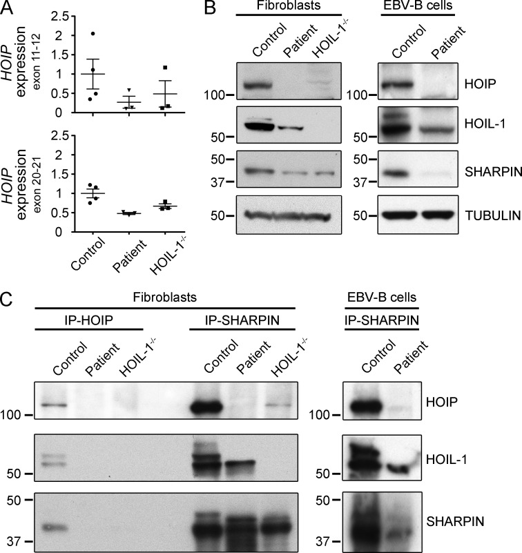
Inherited, complete deficiency of human HOIL-1, a component of the linear ubiquitination chain assembly complex (LUBAC), underlies autoinflammation, infections, and amylopectinosis. We report the clinical description and molecular analysis of a novel inherited disorder of the human LUBAC complex. A patient with multiorgan autoinflammation, combined immunodeficiency, subclinical amylopectinosis, and systemic lymphangiectasia, is homozygous for a mutation in HOIP, the gene encoding the catalytic component of LUBAC. The missense allele (L72P, in the PUB domain) is at least severely hypomorphic, as it impairs HOIP expression and destabilizes the whole LUBAC complex. Linear ubiquitination and NF-κB activation are impaired in the patient's fibroblasts stimulated by IL-1β or TNF. In contrast, the patient's monocytes respond to IL-1β more vigorously than control monocytes. However, the activation and differentiation of the patient's B cells are impaired in response to CD40 engagement. These cellular and clinical phenotypes largely overlap those of HOIL-1-deficient patients. Clinical differences between HOIL-1- and HOIP-mutated patients may result from differences between the mutations, the loci, or other factors. Our findings show that human HOIP is essential for the assembly and function of LUBAC and for various processes governing inflammation and immunity in both hematopoietic and nonhematopoietic cells.


Figures

Figure 1.
Figure 1.
A patient with a homozygous germline HOIP mutation. (A) Amylopectin accumulation in bowel tissue from HOIP-deficient patient. Patient sternocleidomastoid muscular biopsy was stained with hematoxylin and eosin (H&E), or with periodic acid-Schiff stain (PAS) after diastase pretreatment (PASD). A patchy accumulation of PAS+diastase-resistant material in a scattered muscle fibers is shown in patient’ biopsy. Bar, 50 µm. (B) Pedigree of the kindred, showing segregation of the L72P missense mutation. (C) HOIP DNA sequence electropherograms, for controls, the patient and her family. (D) Schematic diagram of the LUBAC complex. The domains identified are depicted as boxes: PNGase/UBA or UBX (PUB), ubiquitin-like (UBL), novel zinc-finger (NZF), really interesting new gene (RING) domain, in-between RING (IBR) domain, zinc finger (ZF). The red star indicates the position of the L72P mutation. Double arrows indicate the domain involved in the interaction between subunits. (E) Amino acid sequences alignment of the PUB domain of HOIP orthologues. The L72 residue is highlighted in red. The hashtags indicate the amino acids conserved in all proteins containing a PUB domain.
Figure 2.
Figure 2.
HOIP deficiency. (A) Relative HOIP mRNA levels in SV40-immortalized fibroblasts from controls, the HOIP-deficient patient (Patient), and HOIL-1–deficient patients (two independent experiments). (B) Expression of LUBAC subunits in control, HOIP-, and HOIL-1–deficient fibroblasts (left) and control and HOIP-deficient EBV-B cells (right) by immunoblotting total cell extracts with the indicated antibodies (two independent experiments). (C) Immunoblot of immunoprecipitated extracts from control, HOIP-, and HOIL-1–deficient fibroblasts (left) and control and HOIP-deficient EBV-B cells (right) with antibodies directed against HOIP (IP-HOIP) or SHARPIN (IP-SHARPIN) and detected with HOIL-1, HOIP, or SHARPIN antibodies (two independent experiments).
Figure 3.
Figure 3.
Impaired TNF- and IL-1β-induced NF-κB activation in the HOIP-deficient patient. (A) Time-course of IL-1β–stimulated (left) or TNF-stimulated (right) SV40-immortalized fibroblasts from control, HOIP-, and HOIL-1–deficient patients, with immunoblotting for phospho-IKK (p-IKK), IKKβ, IκBα, NEMO, and β-tubulin. (B) IL-6 production upon stimulation with TNF and IL-1β in SV-40 fibroblasts from two controls, HOIP- (Patient), HOIL-1–, and NEMO-deficient patients (errors bars indicate the SEM; 3 independent experiments). Unpaired Student’s t tests were performed for the statistical analysis (**, P < 0.01; ***, P < 0.001). (C) Impaired recruitment of NEMO to punctate structures in the patient’s fibroblasts. TNF- or IL-1β–stimulated cells were permeabilized with saponin before fixation, and immunolocalization with anti-NEMO antibody (white) was performed. Nuclei were stained with DAPI. Bar, 10 µm. (D) Absence of linear polyubiquitination in the patients’ fibroblasts upon TNF or IL-1β stimulation. Immunolocalization with anti-linear ubiquitin chains antibody (red) was performed as in C. Nuclei were stained with DAPI. Bars, 10 µm.
Figure 4.
Figure 4.
Complementation of impaired NF-κB activation by reintroduction of the WT HOIP allele. (A) Immunoblot of the three LUBAC subunits in SV-40–immortalized fibroblasts from control, HOIP-deficient (Patient), and HOIL-1–deficient cells expressing empty vector (-) or WT-V5 HOIP constructs (+). (B) Time course of IL-1β−stimulated (top) or TNF-stimulated (bottom) SV-40–immortalized fibroblasts from control, HOIP-deficient (Patient-empty), and HOIP-deficient cells complemented with WT HOIP (Patient-HOIP), with immunoblotting for p-IKK, IKKβ, IκBα, and GAPDH. (C) IL-6 production in SV-40 fibroblasts from control, HOIP- (Patient), or HOIL-1–deficient patients without (-) and with (+) WT HOIP complementation, after stimulation with TNF or IL-1β. Errors bars represent the SEM (five independent experiments). Unpaired Student’s t tests were used for the statistical analysis (**, P < 0.01; ***, P < 0.001; ns, not significant).
Figure 5.
Figure 5.
Impaired B cell activation in the HOIP-deficient patient. (A) Flow cytometry analysis of CD80 expression from stimulated PBMCs. PBMCs left unstimulated or stimulated for 3 d as indicated were stained for CD19 and with either an isotype control (red histograms) or an antibody directed against CD80 (blue histograms). CD80 expression values (CD80 median, isotype median) are indicated under the histograms (one experiment). (B) Flow cytometry analysis of CD27/CD38 expression on gated CD19+ cells, upon stimulation of PBMCs for 7 d with CD40L+IL-21 or with medium alone. The gated populations in the figure correspond to CD38hi and CD27hi plasmablasts (one experiment). (C) Time course of CD40L-stimulated EBV-immortalized B cells from control, HOIP- (patient), and CD40-deficient (CD40−/−) patients, followed by immunoblotting for p-IKK, IKKβ, IκBα, and GAPDH (two independent experiments).
Figure 6.
Figure 6.
Hyperproduction of IL-6 upon IL-1β stimulation in HOIP-deficient monocytes. (A) Production of IL-6 and IL-10 by whole blood cells after 48 h of stimulation with TNF, IL-1β, LPS, and PMA/ionomycin in 32 controls (left), 2 travel controls (middle), and the HOIP-deficient patient (right, in red, 1 experiment). (B) Cytokine production in CD14+ cell subsets, as determined by intracellular FACS staining of stimulated PBMCs. PBMCs from 12 healthy controls (black), the HOIP-deficient patient (red), an HOIL-1–deficient patient (blue), and an IRAK-4–deficient patient (gray) were stimulated with TNF, IL-1β, or LPS. The secretory pathways were concomitantly blocked with monensin and Brefeldin A. 12 h after stimulation, the cells were immunolabeled with antibodies against CD14 and then permeabilized to assess the production of IL-6, IL-1β, IL-8, and MIP-1β by intracellular immunostaining. Labeled cells were analyzed by flow cytometry (3 independent experiments; **, P < 0.01).

References

    1. Abecasis G.R., and Wigginton J.E.. 2005. Handling marker-marker linkage disequilibrium: pedigree analysis with clustered markers. Am. J. Hum. Genet. 77:754–767. 10.1086/497345 - DOI - PMC - PubMed
    1. Abecasis G.R., Cherny S.S., Cookson W.O., and Cardon L.R.. 2002. Merlin—rapid analysis of dense genetic maps using sparse gene flow trees. Nat. Genet. 30:97–101. 10.1038/ng786 - DOI - PubMed
    1. Al-Herz W., Bousfiha A., Casanova J.L., Chatila T., Conley M.E., Cunningham-Rundles C., Etzioni A., Franco J.L., Gaspar H.B., Holland S.M., et al. . 2014. Primary immunodeficiency diseases: an update on the classification from the international union of immunological societies expert committee for primary immunodeficiency. Front. Immunol. 5:162 10.3389/fimmu.2014.00162 - DOI - PMC - PubMed
    1. Alitalo K. 2011. The lymphatic vasculature in disease. Nat. Med. 17:1371–1380. 10.1038/nm.2545 - DOI - PubMed
    1. Allen R.C., Armitage R.J., Conley M.E., Rosenblatt H., Jenkins N.A., Copeland N.G., Bedell M.A., Edelhoff S., Disteche C.M., Simoneaux D.K., et al. . 1993. CD40 ligand gene defects responsible for X-linked hyper-IgM syndrome. Science. 259:990–993. 10.1126/science.7679801 - DOI - PubMed

Publication types

MeSH terms