. 2015 Aug 21;6(24):20084-98.
doi: 10.18632/oncotarget.3907.

A small-molecule inhibitor suppresses the tumor-associated mitochondrial NAD(P)+-dependent malic enzyme (ME2) and induces cellular senescence

Affiliations

A small-molecule inhibitor suppresses the tumor-associated mitochondrial NAD(P)+-dependent malic enzyme (ME2) and induces cellular senescence

Ju-Yi Hsieh et al. Oncotarget. .

Abstract

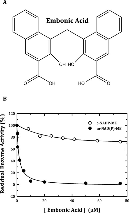
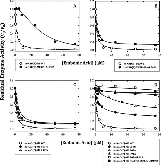
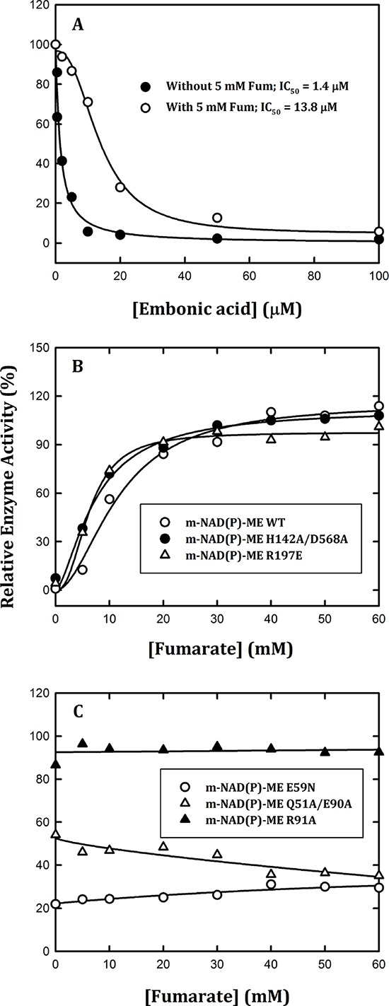
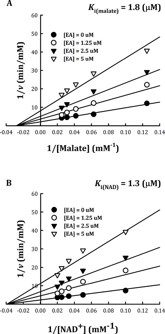
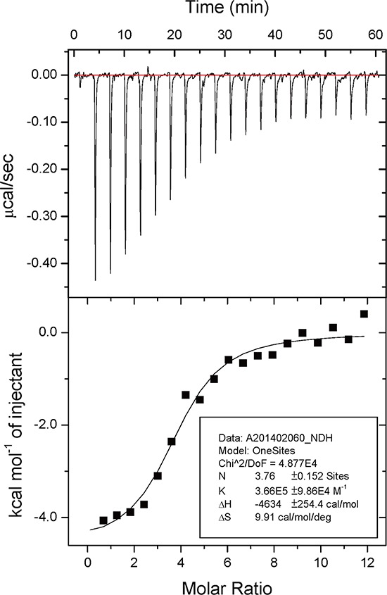
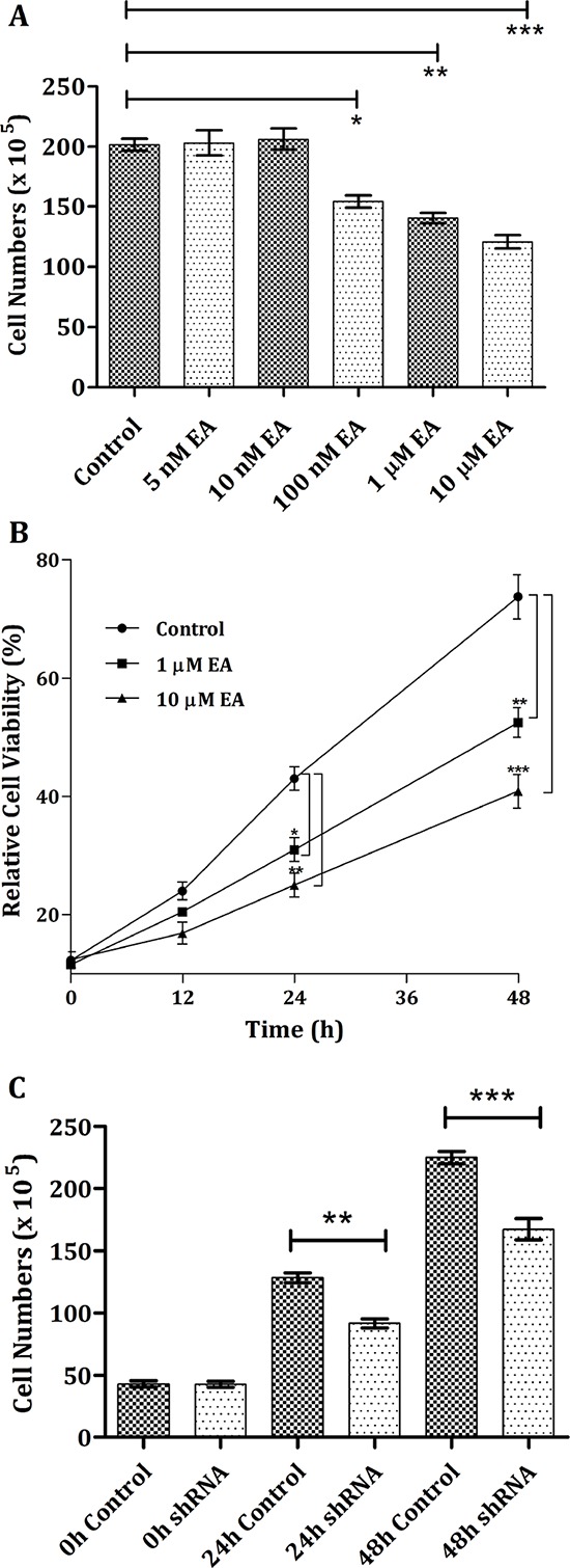
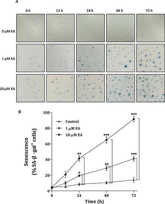
Here, we found a natural compound, embonic acid (EA), that can specifically inhibit the enzymatic activity of mitochondrial NAD(P)+-dependent malic enzyme (m-NAD(P)-ME, ME2) either in vitro or in vivo. The in vitro IC50 value of EA for m-NAD(P)-ME was 1.4 ± 0.4 μM. Mutagenesis and binding studies revealed that the putative binding site of EA on m-NAD(P)-ME is located at the fumarate binding site or at the dimer interface near the site. Inhibition studies reveal that EA displayed a non-competitive inhibition pattern, which demonstrated that the binding site of EA was distinct from the active site of the enzyme. Therefore, EA is thought to be an allosteric inhibitor of m-NAD(P)-ME. Both EA treatment and knockdown of m-NAD(P)-ME by shRNA inhibited the growth of H1299 cancer cells. The protein expression and mRNA synthesis of m-NAD(P)-ME in H1299 cells were not influenced by EA, suggesting that the EA-inhibited H1299 cell growth occurs through the suppression of in vivo m-NAD(P)-ME activity EA treatment further induced the cellular senescence of H1299 cells. However, down-regulation of the enzyme-induced cellular senescence was not through p53. Therefore, the EA-evoked senescence of H1299 cells may occur directly through the inhibition of ME2 or a p53-independent pathway.

Keywords: allosteric inhibitor; cellular senescence; mutagenesis analysis; non-competitive inhibition; selective inhibitor.


Figures

Figure 1
Figure 1. Chemical structure of embonic acid (EA) and inhibitory effect of EA on human m-NAD(P)-ME and c-NADP-ME
A. Chemical structure of EA. B. The assay mixture contained 40 mM malate, 10 mM MgCl2, 2 mM NAD+ or NADP+ and 50 mM Tris-HCl (pH 7.5) with various concentrations of EA. The concentrations of EA ranged from 0 to 75 μM. Closed circles, m-NAD(P)-ME; open circles, c-NADP-ME.
Figure 2
Figure 2. Inhibitory effects of embonic acid (EA) on wild-type and mutant m-NAD(P)-ME
The assay mixture contained 40 mM malate, 10 mM MgCl2, 2 mM NAD+ and 50 mM Tris-HCl (pH 7.5) with various concentrations of EA that ranged from 0 to 75 μM. A. m-NAD(P)-ME WT and the Q51A/E90A dimer interface mutant; B. m-NAD(P)-ME WT and the H142A/D568A tetramer interface mutant; C. m-NAD(P)-ME WT and the R197E and R542V exo site mutants; D. m-NAD(P)-ME WT and the E59N, R67A, R91A, R67A/R91A and K57S/E59N/K73E/D102S fumarate site mutants.
Figure 3
Figure 3. Effects of fumarate on the reversible inhibition of human m-NAD(P)-ME enzyme activity by EA
The assay mixture contained 40 mM malate, 10 mM MgCl2, 2 mM NAD+ and 50 mM Tris-HCl (pH 7.5). A. m-NAD(P)-ME was pre-incubated with 5 mM fumarate and then assayed with various concentrations of EA; B. m-NAD(P)-ME WT (open circles), H142A/D568A (closed circles), and R197E (open triangles) were pre-incubated with 40 μM EA and then assayed with various concentrations of fumarate; C. E59N (open circles), Q51A/E90A (closed circles), and R91A (open triangles) were pre-incubated with 40 μM EA and then assayed with various concentrations of fumarate.
Figure 4
Figure 4. Non-competitive inhibition of human m-NAD(P)-ME by embonic acid (EA)
Human m-NAD(P)-ME activities were measured using different concentrations of A. malate or B. NAD+ with various concentrations of EA; the concentrations of EA were 0, 1.25, 2.5 and 5 μM from the bottom to top.
Figure 5
Figure 5. Isothermal titration calorimetry data of embonic acid (EA) against m-NAD(P)-ME
Human m-NAD(P)-ME (70 μM, 30 mM HEPES, pH 7.4) was titrated with EA (10 mM) at 298K. The upper panel shows the raw data curve, and the lower panel shows the fitted integrated ITC data curve. The data were fitted with the “ONESites” model of the MicroCal (Northampton, MA) version of ORIGIN 7.0.
Figure 6
Figure 6. Cell growth inhibition of H1299 cancer cells through the addition of embonic acid (EA)
A. The dosage performance was examined by treating H1299 cells with vehicle (control), 5, 10, 100 nM, 1 or 10 μM EA for 24 h. B. To determine the time dependence, cells were treated with 0, (control), 1 or 10 μM EA for 0, 12, 24, 36 or 48 h. C. H1299 cells were pretreated with shLuc (control) or ME2 shRNA for 0, 24 or 48 h. Total cells were harvested, and cell numbers were calculated and determined at indicated time and dose points. *P < 0.05, **P < 0.01 and ***P < 0.001.
Figure 7
Figure 7. Senescence of H1299 cancer cells induced by embonic acid (EA)
The dose and time dependencies were determined by treating H1299 cells with different concentrations of EA (0, 1 and 10 μM) for different amounts of time (0, 12, 24, 48 and 72 h), respectively. A. Cells were stained with 5-bromo-4-chloro-3-indolyl β-D-galactoside (X-Gal) and detected using the senescence-associated β-galactosidase (SA-β-gal) assay. The image was analyzed by light microscope. B. The percentages of SA-β-gal+ cells were calculated by the number of positively cells per 1,000 cells counted in ten random fields. *P < 0.05, **P < 0.01 and ***P < 0.001.

Similar articles

Cited by

References

    1. Zheng B, Fisher DE. Metabolic Vulnerability in Melanoma: A ME2 (Me Too) Story. J Invest Dermatol. 2015;135:657–659. - PMC - PubMed
    1. Frenkel R. Regulation and physiological function of malic enzyme. Curr Top Cellu Regul. 1975;9:157–181. - PubMed
    1. Sanz N, Díez-Fernández C, Valverde AM, Lorenzo M, Benito M, Cascales M. Malic enzyme and glucose 6-phosphate dehydrogenase gene expression increases in rat liver cirrhogenesis. Br J Cancer. 1997;75:487–492. - PMC - PubMed
    1. Chang GG, Tong L. Structure and function of malic enzymes, a new class of oxidative decarboxylases. Biochemistry. 2003;42:12721–12733. - PubMed
    1. Loeber G, Infante AA, maurer-Fogy I, Krystek E, Dworkin MB. Human NAD+-dependent mitochondrial malic enzyme. J Biol Chem. 1991;266:3016–3021. - PubMed

Publication types

MeSH terms