. 2015 Jun 30;6(18):16304-20.
doi: 10.18632/oncotarget.4059.

Vitamin D3-dependent VDR signaling delays ron-mediated breast tumorigenesis through suppression of β-catenin activity

Affiliations

Vitamin D3-dependent VDR signaling delays ron-mediated breast tumorigenesis through suppression of β-catenin activity

Abby L Johnson et al. Oncotarget. .

Abstract

The Ron receptor is upregulated in human breast cancers and correlates with enhanced metastasis and reduced patient survival. Ron overexpression drives mammary tumorigenesis through direct β-catenin activation and augmented tumor cell proliferation, migration and invasion. Ron and β-catenin are also coordinately elevated in breast cancers. The vitamin D receptor (VDR) antagonizes β-catenin signaling. Herein, we examined mammary tumor onset and progression using a Ron-driven murine model of breast tumorigenesis crossed with VDR deficient mice. VDR ablation accelerated mammary tumor onset and led to tumors that exhibited a desmoplastic phenotype and enhanced metastases. Tumor levels of active β-catenin were markedly increased in the absence of VDR. In vitro, VDR activation in breast cancer cells reduced β-catenin activation and transcriptional activity leading to elevated expression of the extracellular Wnt inhibitor dickkopf-related protein 1, and a reduction in the interaction of β-catenin with the cyclin D1 promoter. Expression of a stabilized form or β-catenin ablated the protective effects of VDR activation.Collectively, these studies delineate a protective role for VDR signaling in Ron-induced mammary tumorigenesis through disruption of β-catenin activation.

Keywords: breast cancer; ron; vitamin D3 receptor; β-catenin.


Conflict of interest statement

CONFLICTS OF INTEREST

There is no conflict of interest.

Figures
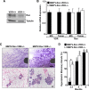
Figure 1
Figure 1. VDR signaling delays Ron-mediated mammary gland hyperplasia
A. Western blot of mammary lysates from MMTV-Ron VDR+/+ and VDR−/− mice depicting Ron expression levels. B. qRT-PCR analysis of Ron and VDR mRNA expression in mammary glands (MG) and tumors from MMTV-Ron VDR+/+ and VDR−/− mice. Data represent mean values from three independent experiments ± SE. C. Representative mammary whole mounts (upper panels) and H&E-stained tissue sections (lower panels) from 10 week-old MMTV-Ron VDR+/+ and VDR−/− mammary glands (n = 8-11). D. Incidence of mice with hyperplastic mammary glands at 2.5, 4 and 6 months of age from MMTV-Ron VDR+/+ and VDR−/− animals. *P < 0.05.
Figure 2
Figure 2. VDR signaling delays mammary tumor formation and alters disease progression in MMTV-Ron mice
A. Mammary tumor kinetics from MMTV-Ron VDR+/+, VDR+/−, and VDR−/− mice. Between 22 and 33 female mice per genotype were examined temporally for palpable tumor formation. The percent tumor-free mice is plotted as a function of age and is statistically significant with complete VDR ablation compared to VDR haploinsufficiency and VDR wild type mice. Inset: The mean time-to-tumor onset was plotted for each genotype. B. Representative mammary glands (upper panels) and tumors (lower panels) from 8 month-old MMTV-Ron VDR+/+ and VDR−/− mice showing enhanced ductal dilation and desmoplasia with VDR ablation (n = 9-11 per genotype). C. Representative immunohistochemical staining for Ron in lung (upper panels) and liver (lower panels) sections from 10 month-old MMTV-Ron VDR+/+ and VDR−/− mice showing more metastases with loss of VDR (n = 5-6 per genotype). D. The average number of metastases per lung (n = 24-25) and liver (n = 14-19) per genotype as assessed by histological analysis is plotted. Data represent mean values ± SE. *P < 0.05.
Figure 3
Figure 3. Enhanced downstream β-catenin signaling in Ron-mediated mammary tumorigenesis with VDR ablation
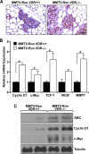
A. Representative immunohistochemical staining for β-catenin in MMTV-Ron VDR+/+ and VDR−/− mammary glands from 4 month-old mice demonstrating enhanced expression in the absence of VDR (n = 6 per genotype). B. qRT-PCR mRNA expression of β-catenin target genes Cyclin D1, c-Myc, TCF-7, VEGF, and MMP7 in mammary tumors from 8 month-old MMTV-Ron VDR+/+ and VDR−/− mice. Data represent mean values from three independent experiments ± SE. C. Western analysis demonstrating enhanced expression of active β-catenin (ABC) levels in two representative MMTV-Ron VDR−/− tumor lysates compared to two MMTV-Ron VDR+/+ tumors that correlates with increased expression of β-catenin target genes cyclin D1 and c-Myc. *P < 0.05.
Figure 4
Figure 4. Ron receptor status determines epithelial cell sensitivity to vitamin D3-mediated growth inhibition and delays in migration and invasion
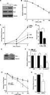
A. Western analysis of levels of VDR and Ron ± 100 nM 1,25D3 in R7 cells. B. R7 cells were treated with the designated concentrations of 1,25D3 for 72 hours and cell viability/number was determined by crystal violet assays. Data are normalized to vehicle treated cells set at 1 and represent mean values from three independent experiments ± SE. C. R7 cells treated with the designated concentrations of 1,25D3 starting at the 0 hour time point were examined in scratch assays for the percent of gap closure after 4 and 8 hours. Data represent mean values from three independent experiments ± SE. D. Matrigel invasion assay with R7 cells in the presence or absence of 100 nM 1,25D3. Data represent mean values from four independent experiments ± SE. E. Western blot demonstrating the reduction in Ron expression in T47D cells stably transfected with a shRon lentivirus. F. qRT-PCR analysis of VDR, Ron and β-catenin mRNA expression in control T47D cells (shNT) and Ron knockdown T47D cells (shRon). Data represent mean values from three independent experiments ± SE. G. MTT assays of T47D control (shNT) and shRon cells treated with the designated concentrations of 1,25D3 for 48 hours. Data represent mean values from 3-4 independent experiments per cell line ± SE. H. Scratch assays in T47D control (shNT) and shRon cells treated with 100 nM 1,25D3 for 24 hours. Data represent mean values normalized to the vehicle-treated control group of each cell type and are from three independent experiments ± SE. *P < 0.05.
Figure 5
Figure 5. Vitamin D3-dependent VDR signaling reduces β-catenin transcriptional activity
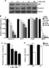
A. Western analysis demonstrating a dose-dependent reduction in active β-catenin (ABC) levels in R7 cells treated with the designated concentrations of 1,25D3 for 48 hours, but no change in total β-catenin. B. qRT-PCR analysis of β-catenin target genes cyclin D1, TCF-7, VEGF, and MMP7 in R7 cells treated with the designated concentrations of 1,25D3 for 72 hours. Data represent mean values from three independent experiments ± SE. C. Dual luciferase assays in R7 cells co-transfected with Topflash and pRLTX (expressing Renilla) plasmids for 48 hours, then treated with the designated concentrations of 1,25D3 for 24 hours. Luciferase activities were normalized for transfection efficiency to Renilla activity. Data represent mean values from three independent experiments ± SE. D. Crystal violet staining of R7 cells transfected with a stabilized form of β-catenin (ΔN) or empty vector and treated with 100 nM 1,25D3 or vehicle control (EtOH). Data represent mean values from four independent experiments ± SE. *P < 0.05.
Figure 6
Figure 6. Vitamin D3-dependent VDR signaling induces DKK-1 expression and binds to β-catenin to disrupt interaction at consensus sequences within promoters of TCF/LEF target genes
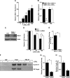
A. qRT-PCR mRNA expression of DKK-1 in R7 cells treated with the designated concentrations of 1,25D3 for 72 hours. Data represent mean values from three independent experiments ± SE. B. qRT-PCR mRNA expression of DKK-1 in tumors and mammary glands (MG) from 8 month-old MMTV-Ron VDR+/+ and VDR−/− mice demonstrating a reduction in DKK-1 levels in tumors with loss of VDR. Data represent mean values from three independent experiments ± SE. C. Western blot demonstrating loss of DKK-1 protein expression with siRNA-mediated silencing in R7 cells. D. R7 cells transfected with siRNA against DKK-1 were treated with the designated concentrations of 1,25D3 for 72 hours and cell viability/number was determined by crystal violet assays. Data are normalized to the respective vehicle treated cells set at 1 and represent mean values from two independent experiments performed in quadruplicate ± SE. E. Chromatin immunoprecipitation (ChIP) assays with a mouse IgG isotype control and an anti-active β-catenin (ABC) antibody. ChIP-ABC quantitative real time PCR (qRT-PCR) analysis of R7 cells treated with 100 nM 1,25D3 for 72 hours. The graph shows qRT-PCR on DNA purified from ChIP-ABC, using primers designed to the LEF-1 binding sequence within the mouse cyclin D1 promoter and relative to the respective input controls. Vitamin D3 treatment significantly reduces enrichment compared to the vehicle control (EtOH). Data represent mean values from three independent experiments ± SE. F. Representative agarose gel from ChIP-ABC PCR showing reduced cyclin D1 promoter enrichment with vitamin D3 treatment in R7 cells. Negative control primers designed to a site 4000 bp upstream of the LEF-1 binding sequence within the cyclin D1 promoter (Off-Target) verify specificity of cyclin D1 primers to the sheared DNA product. G. Densitometry analysis of PCR products from three separate ChIP-ABC experiments supporting loss of ABC interaction with the cyclin D1 promoter. Error bars represent SE. H. Re-ChIP qRT-PCR analysis of R7 cells treated with 100 nM 1,25D3 for 72 hours and sequentially immunoprecipitated with anti-ABC then anti-VDR antibodies. The graph shows qRT-PCR of DNA purified from ChIP-ABC-VDR, using primers designed to the LEF-1 binding sequence within the mouse cyclin D1 promoter, and showing less interaction of the ABC-VDR complex at the cyclin D1 promoter. Data represent the relative mean CT values from two experiments ± standard deviation. *P < 0.05.
Figure 7
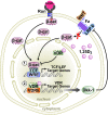
Figure 7. A working model of VDR signaling in Ron-mediated breast cancer
Binding of the Ron ligand, HGFL, to Ron induces β-catenin activation promoting breast cancer growth and metastasis when aberrantly expressed. VDR signaling in breast cancer cells antagonizes β-catenin activity, delaying tumor initiation and progression through two mechanisms: 1) Vitamin D3-bound VDR can directly interact with β-catenin, preventing its association with TCF/LEF transcription factors thus inhibiting transcriptional activity; and, 2) ligand-dependent VDR signaling can regulate the transcription of various VDR target genes. One such VDR target includes DKK-1, which inhibits canonical Wnt signaling, thereby reducing cytosolic accumulation of β-catenin and nuclear localization for transcriptional activity.

Similar articles

Cited by

References

    1. Maggiora P, Marchio S, Stella MC, Giai M, Belfiore A, De Bortoli M, Di Renzo MF, Costantino A, Sismondi P, Comoglio PM. Overexpression of the RON gene in human breast carcinoma. Oncogene. 1998;16:2927–2933. - PubMed
    1. Lee WY, Chen HH, Chow NH, Su WC, Lin PW, Guo HR. Prognostic significance of co-expression of RON and MET receptors in node-negative breast cancer patients. Clin Cancer Res. 2005;11:2222–2228. - PubMed
    1. Welm AL, Sneddon JB, Taylor C, Nuyten DS, van de Vijver MJ, Hasegawa BH, Bishop JM. The macrophage-stimulating protein pathway promotes metastasis in a mouse model for breast cancer and predicts poor prognosis in humans. Proceedings of the National Academy of Sciences of the United States of America. 2007;104:7570–7575. - PMC - PubMed
    1. Kretschmann KL, Eyob H, Buys SS, Welm AL. The macrophage stimulating protein/Ron pathway as a potential therapeutic target to impede multiple mechanisms involved in breast cancer progression. Current drug targets. 2010;11:1157–1168. - PubMed
    1. Gaudino G, Follenzi A, Naldini L, Collesi C, Santoro M, Gallo KA, Godowski PJ, Comoglio PM. RON is a heterodimeric tyrosine kinase receptor activated by the HGF homologue MSP. The EMBO journal. 1994;13:3524–3532. - PMC - PubMed

Publication types

MeSH terms

LinkOut - more resources