. 2015 Jul 1;107(1):66-77.
doi: 10.1093/cvr/cvv152. Epub 2015 May 25.

Oestrogen enhances cardiotoxicity induced by Sunitinib by regulation of drug transport and metabolism

Affiliations

Oestrogen enhances cardiotoxicity induced by Sunitinib by regulation of drug transport and metabolism

Pamela Ann Harvey et al. Cardiovasc Res. .

Abstract

Aims: To define the molecular mechanisms of cardiotoxicity induced by Sunitinib and to identify the role of biological sex in modulating toxicity.

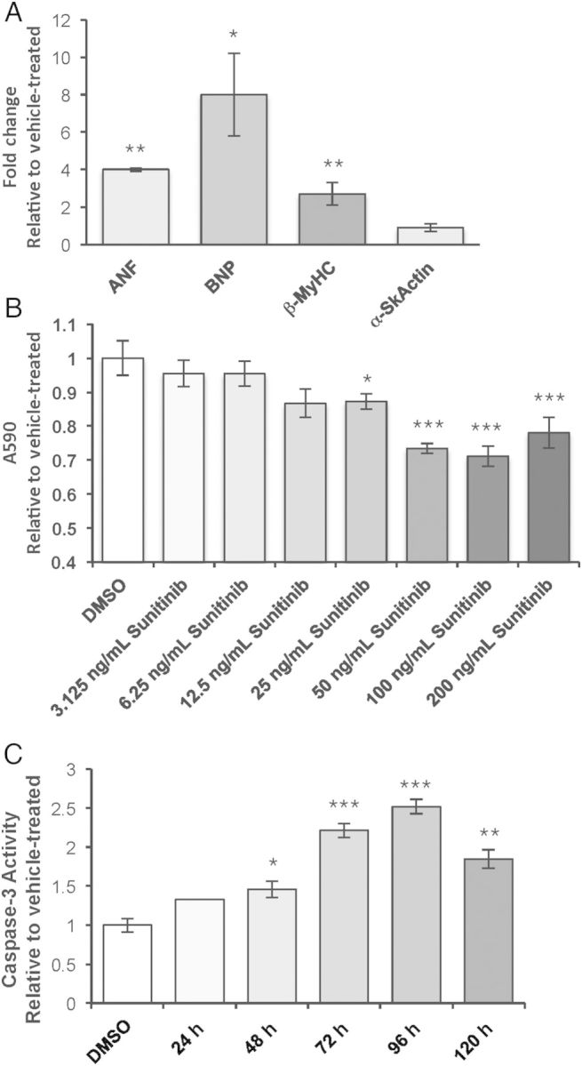
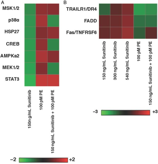
Methods and results: Exposure of isolated cardiomyocytes to plasma-relevant concentrations of Sunitinib and other tyrosine kinase inhibitors produces a broad spectrum of abnormalities and cell death via apoptosis downstream of sexually dimorphic kinase inhibition. Phosphorylation of protein kinase C and phospholipase γ abrogates these effects for most tyrosine kinase inhibitors tested. Female sex and estradiol cause increased cardiotoxicity, which is mediated by reduced expression of a drug efflux transporter and a metabolic enzyme. Female but not male mice exposed to a 28-day course of oral Sunitinib exhibit similar abnormalities as well as functional deficits and their hearts exhibit differential expression of genes responsible for transport and metabolism of Sunitinib.

Conclusion: We identify the specific pathways affected by tyrosine kinase inhibitors in mammalian cardiomyocytes, interactions with biological sex, and a role for oestrogen in modulating drug efflux and metabolism. These findings represent a critical step toward reducing the incidence of cardiotoxicity with tyrosine kinase inhibitor chemotherapeutics.

Keywords: Cardiotoxicity; Cytochrome P450; Multidrug-resistance-1; Oestrogen; Sunitinib.


Figures

Figure 1
Figure 1
TKIs induce cardiotoxicity and cell death in NRVMs. (A) Fetal gene expression was measured in NRVMs treated with 150 ng/mL Sunitinib for 36 h using quantitative PCR. Fold change is relative to vehicle-treated. ANF, atrial natriuretic factor; BNP, brain natriuretic peptide; β-MyHC, β-myosin heavy chain; α-SkActin, α-Skeletal actin. n = 4 NRVM preparations analysed separately. Bars indicate the average fold change of the four preparations. Student's t-test, comparing Sunitinib- to vehicle-treated cells. (B) Absorbance at 590 nm (A590) of crystal violet-stained NRVMs treated with vehicle (DMSO), 75 ng/mL, 150 ng/mL, or 300 ng/mL Sunitinib (Sun) for 36 h. n = 6 NRVM preparations analysed separately. Bars indicate the average of the six preparations. One-way ANOVA followed by Tukey's post-hoc test. (C) Fold change in caspase-3 activity measured in lysates from NRVMs treated with 150 ng/mL Sunitinib for 24, 48, 72, 96, or 120 h. n = 3 NRVM preparations of 80–100 neonatal ventricles each. One-way ANOVA followed by Tukey's post-hoc test. For each panel, error bars represent standard error from the mean (SEM). *P < 0.05, **P < 0.01.
Figure 2
Figure 2
Off-target kinase inhibition induced by TKIs (150 ng/mL Sunitinib, 1 μg/mL Erlotinib, 3 μg/mL Imatinib, 100 ng/mL Dasatinib, 1.4 μg/mL Lapatinib, or 500 ng/mL Sorafenib) in NRVMs. (A) Levels of phosphorylation of RTKs in NRVMs treated with plasma-relevant doses of TKIs for 24 h were measured using an antibody array. Data were obtained from a single antibody array using three pooled NRVM preparations (80–100 neonatal ventricles each, 270 ventricle total) with each RTK tested in duplicate. Green indicates decreased phosphorylation and red indicates increased phosphorylation, relative to vehicle-treated NRVMs. (B) Absorbance of NRVMs treated with TKIs for 24 h and stained with crystal violet (A590). n = 4 NRVM preparations (80–100 neonatal ventricles per preparation) analysed separately and averaged. *P < 0.05, **P < 0.01, one-way ANOVA followed by Tukey's post-hoc test.
Figure 3
Figure 3
Phosphorylation of PKC and PLCγ rescues cell loss in NRVMs treated with TKIs. A590 (absorbance of samples stained with crystal violet) was used to measure cell death in NRVMs treated with Sunitinib alone (light grey bar) or co-treated with decreasing concentrations of phenylephrine (PE, dark grey bars) for 24 h (A) with Sunitinib alone (white bar) or combined with the specific protein kinase C activator, phorbol 12-myristate 13-acetate (PMA) (hatched bar), for 24 h. Hatched bars indicate co-treatment with 50 nM PMA (B) or with TKIs alone (white bar) or combined with 2.8 pM PE for 24 h (hatched bars). (C and D) Caspase-3 activity measured in lysates obtained from NRVMs treated with TKIs alone or combined with 2.8 nM PE for 24 h. (A–C) n = 6 NRVM preparations; (D) n = 3 NRVM preparations. Error bars represent SEM. *P < 0.05, **P < 0.01, relative to vehicle using Student's t-test (A and C) or among all groups using ANOVA followed by Tukey's post-hoc test (B and D).
Figure 4
Figure 4
(A) Heat map demonstrating phosphorylation of intracellular signalling molecules in NRVMs treated with Sunitinib, PE, or both for 24 h. (B) Heat map demonstrating levels of protein involved in apoptosis in NRVMs treated for 24 h with increasing doses of Sunitinib (150, 300, 450 ng/mL), 100 pM PE, or 150 ng/mL Sunitinib plus 100 pM PE. (A and B) n = 3 pooled NRVM preparations, 80–100 neonatal ventricles per preparation.
Figure 5
Figure 5
(A) Expression of the fetal gene program (ANF, BNP, βMyHC, SERCA) in female (light grey bars) and male (dark grey bars) ARVMs treated with 150 ng/mL Sunitinib or vehicle (DMSO) for 36 h. n = ventricles from four male and four female rats evaluated individually and averaged, Student's t-test, comparing Sunitinib with vehicle-treated cells. (B) Caspase-3 activity measured using a colorimetric assay in female (light grey bars) and male (dark grey bars) ARVMs treated with Sunitinib or vehicle for 36 h. n = ventricles from four male and four female rats evaluated separately and averaged, Student's t-test, comparing Sunitinib to vehicle-treated. (C) Heat map demonstrating activation states of RTKs in female and male ARVMs treated with Sunitinib, relative to vehicle-treated. n = ventricles from four male and fur female rats. Two RTKs antibody arrays were performed using two pooled male and female ARVM preparations each. Student's t-test, comparing Sunitinib to vehicle-treated cells. For A and B, error bars represent SEM. *P < 0.05, **P < 0.01.
Figure 6
Figure 6
(A) Expression of MDR1 in female (light grey bars) and male ARVMs treated with Sunitinib. MDR1 (B) or CYP1A1 (C) expression in NRVMs treated with Sunitinib or plasma-relevant concentrations of oestrogen (E2) alone or combined with 100 nM E2 or 1 μM E2 (hatched bars) for 24 h. (D) ANF expression in NRVMs treated with Sunitinib alone (white bar) or with E2 or PE (white hatched bars), or with Ellipticine (black bar). Error bars indicate SEM. (A) Student's t-test, compared with Sunitinib treated. n = 4 rats per sex. (B–D) One-way ANOVA followed by Tukey's post-hoc test. n = 4 rats per sex. (A–D) *P < 0.05, **P < 0.01, ***P < 0.001.
Figure 7
Figure 7
Mice receiving Sunitinib exhibit sexually dimorphic functional and molecular cardiotoxicity. (A) Percent fractional shortening (%FS), (B) Left-ventricular anterior wall thickness during systole (LVAW;s) in female (light grey bars) and male (dark grey bars) mice receiving vehicle (open bars) or Sunitinib (hatched bars) for 28 days. (C) Left-ventricular volume during systole (LV Vol;s), (D–F) Expression of fetal genes (D) MDR1 (E) and CYP1A1 (F) in the left ventricles of female and male mice. Female mice, light grey bars; male mice, dark grey bars. (A–C, E and F) two-way ANOVA followed by Tukey's post-hoc test, (D) Student's t-test comparing expression of individual genes between males and females. (A–F) Error bars represent SEM. *P < 0.05, **P < 0.01,***P < 0.001, n = 6–7 mice per sex.

References

    1. Zhang J, Yang PL, Gray NS. Targeting cancer with small molecule kinase inhibitors. Nat Rev Cancer 2009;9:28–39. - PubMed
    1. Chu TF, Rupnick MA, Kerkela R, Dallabrida SM, Zurakowski D, Nguyen L, Woulfe K, Pravda E, Cassiola F, Desai J, George S, Morgan JA, Harris DM, Ismail NS, Chen JH, Schoen FJ, Van den Abbeele AD, Demetri GD, Force T, Chen MH. Cardiotoxicity associated with tyrosine kinase inhibitor sunitinib. Lancet 2007;370:2011–2019. - PMC - PubMed
    1. Hall PH, Harshman LC, Srinivas S, Witteless RM. The frequency and severity of cardiovascular toxicity from targeted therapy in advanced renal cell carcinoma patients. JACC Heart Failure 2013;1:72–78. - PubMed
    1. Telli ML, Witteles RM, Fisher GA, Srinivas S. Cardiotoxicity associated with the cancer therapeutic agent sunitinib malate. Ann Oncol 2008;19:1613–1618. - PubMed
    1. Kerkela R, Grazette L, Yacobi R, Iliescu C, Patten R, Beahm C, Walters B, Shevtsov S, Pesant S, Clubb FJ, Rosenzweig A, Salomon RN, Van Etten RA, Alroy J, Durand JB, Force T. Cardiotoxicity of the cancer therapeutic agent imatinib mesylate. Nat Med 2006;12:908–916. - PubMed

Publication types

MeSH terms