

Extrasynaptic release of GABA and dopamine by retinal dopaminergic neurons
- PMID: 26009765
- PMCID: PMC4455755
- DOI: 10.1098/rstb.2014.0186
Extrasynaptic release of GABA and dopamine by retinal dopaminergic neurons
Abstract
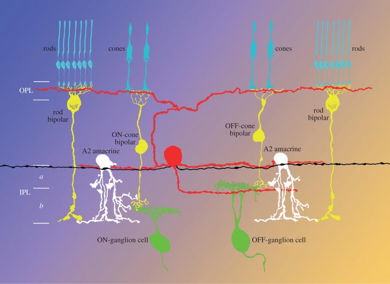
In the mouse retina, dopaminergic amacrine (DA) cells synthesize both dopamine and GABA. Both transmitters are released extrasynaptically and act on neighbouring and distant retinal neurons by volume transmission. In simultaneous recordings of dopamine and GABA release from isolated perikarya of DA cells, a proportion of the events of dopamine and GABA exocytosis were simultaneous, suggesting co-release. In addition, DA cells establish GABAergic synapses onto AII amacrine cells, the neurons that transfer rod bipolar signals to cone bipolars. GABAA but not dopamine receptors are clustered in the postsynaptic membrane. Therefore, dopamine, irrespective of its site of release-synaptic or extrasynaptic-exclusively acts by volume transmission. Dopamine is released upon illumination and sets the gain of retinal neurons for vision in bright light. The GABA released at DA cells' synapses probably prevents signals from the saturated rods from entering the cone pathway when the dark-adapted retina is exposed to bright illumination. The GABA released extrasynaptically by DA and other amacrine cells may set a 'GABAergic tone' in the inner plexiform layer and thus counteract the effects of a spillover of glutamate released at the bipolar cell synapses of adjacent OFF and ON strata, thus preserving segregation of signals between ON and OFF pathways.
Keywords: GABA; dopamine; extrasynaptic release; retina.
© 2015 The Author(s) Published by the Royal Society. All rights reserved.
Figures
References
-
- Witkovsky P, Dearry A. 1991. Functional roles of dopamine in the vertebrate retina. Progr. Retinal Res. 11, 247–292. ( 10.1016/0278-4327(91)90031-V) - DOI
Publication types
MeSH terms
Substances
Grants and funding
LinkOut - more resources
Full Text Sources
Other Literature Sources
Miscellaneous