

Effect of amyloids on the vesicular machinery: implications for somatic neurotransmission
- PMID: 26009766
- PMCID: PMC4455756
- DOI: 10.1098/rstb.2014.0187
Effect of amyloids on the vesicular machinery: implications for somatic neurotransmission
Abstract
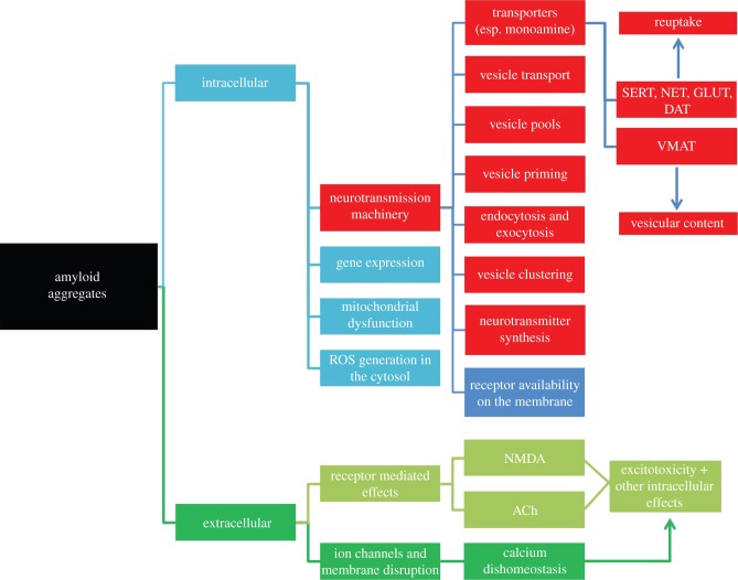
Certain neurodegenerative diseases are thought to be initiated by the aggregation of amyloidogenic proteins. However, the mechanism underlying toxicity remains obscure. Most of the suggested mechanisms are generic in nature and do not directly explain the neuron-type specific lesions observed in many of these diseases. Some recent reports suggest that the toxic aggregates impair the synaptic vesicular machinery. This may lead to an understanding of the neuron-type specificity observed in these diseases. A disruption of the vesicular machinery can also be deleterious for extra-synaptic, especially somatic, neurotransmission (common in serotonergic and dopaminergic systems which are specifically affected in Alzheimer's disease (AD) and Parkinson's disease (PD), respectively), though this relationship has remained unexplored. In this review, we discuss amyloid-induced damage to the neurotransmitter vesicular machinery, with an eye on the possible implications for somatic exocytosis. We argue that the larger size of the system, and the availability of multi-photon microscopy techniques for directly visualizing monoamines, make the somatic exocytosis machinery a more tractable model for understanding the effect of amyloids on all types of vesicular neurotransmission. Indeed, exploring this neglected connection may not just be important, it may be a more fruitful route for understanding AD and PD.
Keywords: Alzheimer's; Parkinson's; monoamine; multiphoton microscopy; neurodegeneration; protein aggregation.
© 2015 The Author(s) Published by the Royal Society. All rights reserved.
Figures
Similar articles
-
Vesicular neurotransmitter transporters as targets for endogenous and exogenous toxic substances.Annu Rev Pharmacol Toxicol. 2008;48:277-301. doi: 10.1146/annurev.pharmtox.46.120604.141146. Annu Rev Pharmacol Toxicol. 2008. PMID: 17883368 Review.
-
The dynamics of somatic exocytosis in monoaminergic neurons.Front Physiol. 2012 Nov 6;3:414. doi: 10.3389/fphys.2012.00414. eCollection 2012. Front Physiol. 2012. PMID: 23133421 Free PMC article.
-
Phthalocyanines as Molecular Scaffolds to Block Disease-Associated Protein Aggregation.Acc Chem Res. 2016 May 17;49(5):801-8. doi: 10.1021/acs.accounts.5b00507. Epub 2016 May 2. Acc Chem Res. 2016. PMID: 27136297 Review.
-
Somatodendritic dopamine release: recent mechanistic insights.Philos Trans R Soc Lond B Biol Sci. 2015 Jul 5;370(1672):20140185. doi: 10.1098/rstb.2014.0185. Philos Trans R Soc Lond B Biol Sci. 2015. PMID: 26009764 Free PMC article. Review.
-
What is the function of neuronal AP-3?Biol Cell. 2007 Jul;99(7):349-61. doi: 10.1042/BC20070029. Biol Cell. 2007. PMID: 17567262 Review.
Cited by
-
Chronic Cerebral Hypoperfusion Aggravates Parkinson's Disease Dementia-Like Symptoms and Pathology in 6-OHDA-Lesioned Rat through Interfering with Sphingolipid Metabolism.Oxid Med Cell Longev. 2022 Aug 8;2022:5392966. doi: 10.1155/2022/5392966. eCollection 2022. Oxid Med Cell Longev. 2022. PMID: 35979400 Free PMC article.
-
Release of chemical transmitters from cell bodies and dendrites of nerve cells.Philos Trans R Soc Lond B Biol Sci. 2015 Jul 5;370(1672):20140181. doi: 10.1098/rstb.2014.0181. Philos Trans R Soc Lond B Biol Sci. 2015. PMID: 26009760 Free PMC article.
-
Predicting Amyloidogenic Proteins in the Proteomes of Plants.Int J Mol Sci. 2017 Oct 16;18(10):2155. doi: 10.3390/ijms18102155. Int J Mol Sci. 2017. PMID: 29035294 Free PMC article.
-
Dopamine transporter forms stable dimers in the live cell plasma membrane in a phosphatidylinositol 4,5-bisphosphate-independent manner.J Biol Chem. 2019 Apr 5;294(14):5632-5642. doi: 10.1074/jbc.RA118.006178. Epub 2019 Jan 31. J Biol Chem. 2019. PMID: 30705091 Free PMC article.
References
-
- Gyure KA, Durham R, Stewart WF, Smialek JE, Troncoso JC. 2001. Intraneuronal aβ-amyloid precedes development of amyloid plaques in Down syndrome. Arch. Pathol. Lab. Med. 125, 489–492. - PubMed
Publication types
MeSH terms
Substances
LinkOut - more resources
Full Text Sources
Other Literature Sources
Medical
