

The structure and function of 'active zone material' at synapses
- PMID: 26009768
- PMCID: PMC4455758
- DOI: 10.1098/rstb.2014.0189
The structure and function of 'active zone material' at synapses
Abstract
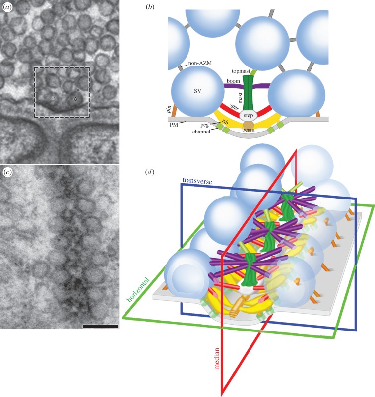
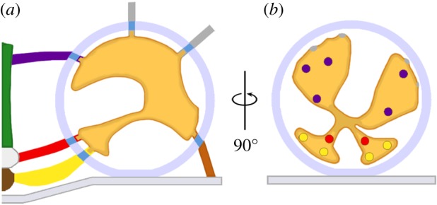
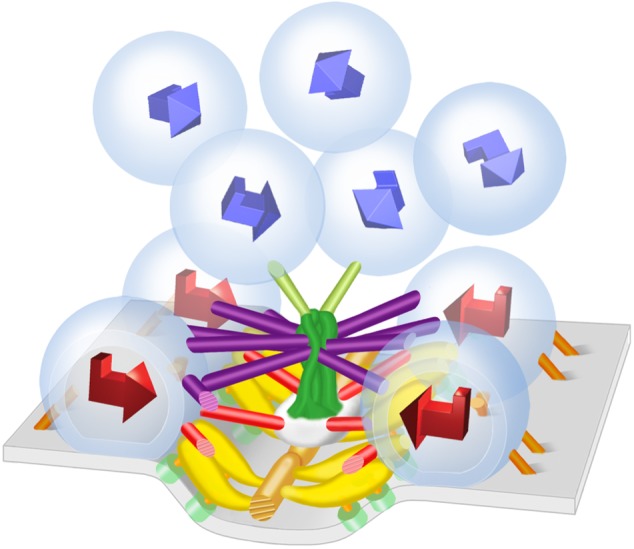
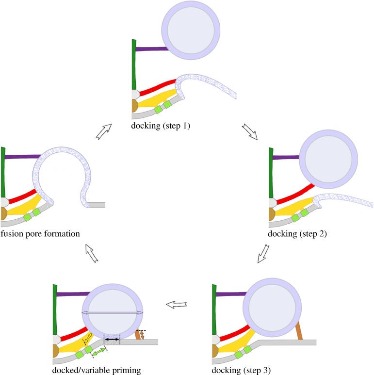
The docking of synaptic vesicles on the presynaptic membrane and their priming for fusion with it to mediate synaptic transmission of nerve impulses typically occur at structurally specialized regions on the membrane called active zones. Stable components of active zones include aggregates of macromolecules, 'active zone material' (AZM), attached to the presynaptic membrane, and aggregates of Ca(2+)-channels in the membrane, through which Ca(2+) enters the cytosol to trigger impulse-evoked vesicle fusion with the presynaptic membrane by interacting with Ca(2+)-sensors on the vesicles. This laboratory has used electron tomography to study, at macromolecular spatial resolution, the structure and function of AZM at the simply arranged active zones of axon terminals at frog neuromuscular junctions. The results support the conclusion that AZM directs the docking and priming of synaptic vesicles and essential positioning of Ca(2+)-channels relative to the vesicles' Ca(2+)-sensors. Here we review the findings and comment on their applicability to understanding mechanisms of docking, priming and Ca(2+)-triggering at other synapses, where the arrangement of active zone components differs.
Keywords: active zone material; docking; electron tomography; priming; synaptic transmission; synaptic vesicle.
© 2015 The Author(s) Published by the Royal Society. All rights reserved.
Figures
Similar articles
-
Regulation of synaptic vesicle docking by different classes of macromolecules in active zone material.PLoS One. 2012;7(3):e33333. doi: 10.1371/journal.pone.0033333. Epub 2012 Mar 16. PLoS One. 2012. PMID: 22438915 Free PMC article.
-
Alignment of synaptic vesicle macromolecules with the macromolecules in active zone material that direct vesicle docking.PLoS One. 2013 Jul 22;8(7):e69410. doi: 10.1371/journal.pone.0069410. Print 2013. PLoS One. 2013. PMID: 23894473 Free PMC article.
-
Macromolecular connections of active zone material to docked synaptic vesicles and presynaptic membrane at neuromuscular junctions of mouse.J Comp Neurol. 2009 Apr 10;513(5):457-68. doi: 10.1002/cne.21975. J Comp Neurol. 2009. PMID: 19226520 Free PMC article.
-
Synaptic Vesicles Having Large Contact Areas with the Presynaptic Membrane are Preferentially Hemifused at Active Zones of Frog Neuromuscular Junctions Fixed during Synaptic Activity.Int J Mol Sci. 2019 May 31;20(11):2692. doi: 10.3390/ijms20112692. Int J Mol Sci. 2019. PMID: 31159267 Free PMC article. Review.
-
Ca(2+) channels and transmitter release at the active zone.Cell Calcium. 2012 Sep-Oct;52(3-4):199-207. doi: 10.1016/j.ceca.2012.04.011. Epub 2012 Jun 8. Cell Calcium. 2012. PMID: 22682961 Review.
Cited by
-
Release of chemical transmitters from cell bodies and dendrites of nerve cells.Philos Trans R Soc Lond B Biol Sci. 2015 Jul 5;370(1672):20140181. doi: 10.1098/rstb.2014.0181. Philos Trans R Soc Lond B Biol Sci. 2015. PMID: 26009760 Free PMC article.
-
The Structure of Human Neuromuscular Junctions: Some Unanswered Molecular Questions.Int J Mol Sci. 2017 Oct 19;18(10):2183. doi: 10.3390/ijms18102183. Int J Mol Sci. 2017. PMID: 29048368 Free PMC article. Review.
-
A Comparison of the Primary Sensory Neurons Used in Olfaction and Vision.Front Cell Neurosci. 2020 Nov 5;14:595523. doi: 10.3389/fncel.2020.595523. eCollection 2020. Front Cell Neurosci. 2020. PMID: 33250719 Free PMC article. Review.
-
Oxidative Stress, Synaptic Dysfunction, and Alzheimer's Disease.J Alzheimers Dis. 2017;57(4):1105-1121. doi: 10.3233/JAD-161088. J Alzheimers Dis. 2017. PMID: 28059794 Free PMC article. Review.
-
Presynaptic mechanisms controlling calcium-triggered transmitter release at the neuromuscular junction.Curr Opin Physiol. 2018 Aug;4:15-24. doi: 10.1016/j.cophys.2018.03.004. Epub 2018 Mar 17. Curr Opin Physiol. 2018. PMID: 30272045 Free PMC article.
References
Publication types
MeSH terms
Substances
Grants and funding
LinkOut - more resources
Full Text Sources
Other Literature Sources
Miscellaneous
