. 2015 Aug 21;6(24):20723-39.
doi: 10.18632/oncotarget.3978.

Hypoxia promotes colon cancer dissemination through up-regulation of cell migration-inducing protein (CEMIP)

Affiliations

Hypoxia promotes colon cancer dissemination through up-regulation of cell migration-inducing protein (CEMIP)

Nikki A Evensen et al. Oncotarget. .

Abstract

Hypoxic stress drives cancer progression by causing a transcriptional reprogramming. Recently, KIAA1199 was discovered to be a cell-migration inducing protein (renamed CEMIP) that is upregulated in human cancers. However, the mechanism of induction of CEMIP in cancer was hitherto unknown. Here we demonstrate that hypoxia induces CEMIP expression leading to enhanced cell migration. Immunohistochemistry of human colon cancer tissues revealed that CEMIP is upregulated in cancer cells located at the invasive front or in the submucosa. CEMIP localization inversely correlated with E-cadherin expression, which is characteristic of the epithelial-to-mesenchymal transition. Mechanistically, hypoxia-inducible-factor-2α (HIF-2α), but not HIF-1α binds directly to the hypoxia response element within the CEMIP promoter region resulting in increased CEMIP expression. Functional characterization reveals that CEMIP is a downstream effector of HIF-2α-mediated cell migration. Expression of CEMIP was demonstrated to negatively correlate with the expression of Jarid1A, a histone demethylase that removes methyl groups from H3K4me3 (an activation marker for transcription), resulting in altered gene repression. Low oxygen tension inhibits the function of Jarid1A, leading to increased presence of H3K4me3 within the CEMIP promoter. These results provide insight into the upregulation of CEMIP within cancer and can lead to novel treatment strategies targeting this cancer cell migration-promoting gene.

Keywords: HIF-2α; KIAA1199/CEMIP; invasion; migration.


Figures
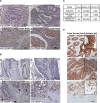
Figure 1
Figure 1. Upregulation of CEMIP in human invasive and metastatic colon cancer
A and B. A human colon cancer tissue microarray (US Biomax CO1005) containing tumor adjacent normal tissue (A-a), adenocarcinomas (A-b), and adenocarcinomas metastasized to lymph nodes (A-c & d), and individual human colon adenocarcinoma specimens containing cancer adjacent normal tissues (B-a), primary tumor (B-b), invasive front (B-c), and invaded tumor cells in submucosa (B-d) were examined by immunohistochemistry (IHC) with an anti-CEMIP antibody. Representative images of tissue types and staining intensities of colon cancer tissue array and tissue samples are shown. Bar = 100 μm. C. Total number of individual specimens from human colon adenoma, adenocarcinoma and adjacent normal tissue examined by IHC for CEMIP expression. D. Individual human colon adenocarcinoma specimens containing cancer adjacent normal tissues (D-a), primary tumor (D-b), and invaded tumor cells in submucosa (D-c) were examined by IHC with an anti-E-cadherin antibody. Representative images are shown. Bar = 100 μm. The staining for CEMIP in panel B and E-cadherin in panel D were from the same case. Inserts represent enlarged images of representative positive cells.
Figure 2
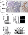
Figure 2. Hypoxia induces CEMIP expression in human colon cancer
A. Hypoxia induces HIF2-α and CA9 in SW480 cells. Total cell lysates from SW480 cells cultured under either normoxia or hypoxia for 48 hours were examined by Western blotting using antibodies as indicated. Actin was used as loading control for Western blotting. B. Human colon adenocarcinoma specimens were stained with anti-CA9 and anti-CEMIP antibodies separately. Representative images from the same case are shown. C. Total RNA and lysates isolated from SW480 and HCT-116 cells were analyzed by real time RT-PCR using CEMIP-specific primers and Western blotting using anti-CEMIP antibody. The mRNA expression was normalized using housekeeping gene HPRT-1 and tubulin was used as loading control for Western blotting. D. Cell migratory ability of SW480 and HCT-116 cells was examined by a Transwell chamber migration assay. Migrated cells were stained by Hoechst. Representative fields are shown in the right panel. E. Total RNA and lysates were analyzed by real time RT-PCR and Western blotting in SW480 cells cultured under normoxic (21% O2) or hypoxic (1% O2) conditions for indicated times. F. Western blot analysis of whole cells lysates from MCF-7 cells cultured under normoxic or hypoxic conditions for indicated times. MCF-7 cells transfected with HIF-1αmu or HIF-2αmu cDNAs were used as positive controls. Tubulin was used as a loading control.
Figure 3
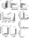
Figure 3. HIF-2α directly induces CEMIP transcription
A. Dual luciferase reporter assay using lysates from HCT-116 and HeLa cells transfected with pGL3 basic (control), wild-type pro-1.4 kb CEMIP, or ΔHRE pro-1.4 kb CEMIP promoter-luciferase reporter cDNAs cultured under normoxia (nor) or hypoxia (hyp) for 48 hours. Renilla luciferase was used as a normalization control. B. Dual luciferase reporter assay using lysates from COS-1 cells transfected with CEMIP promoter-luciferase reporter cDNA along with increasing amounts of vector control or HIF-α mutant cDNAs as indicated. Renilla luciferase was used as a normalization control. C. Dual luciferase reporter assay using lysates from SW480 and HeLa cells co-transfected with indicated cDNAs. Renilla luciferase was used as a normalization control. D. A chromatin immunoprecipitation (ChIP) assay using HeLa cells either transfected with indicated cDNAs or cultured under normoxia or hypoxia for 48 hours. An anti-HIF-2α-specific antibody or an IgG control antibody was used for the ChIP. Primers spanning the HRE within the CEMIP promoter region were used for quantitative PCR. Primers that span the outside of the hypoxia response element within the CEMIP promoter were used as negative control.
Figure 4
Figure 4. HIF-2α leads to increased cell migration that is dependent on upregulated CEMIP
A. Left Panel: Real time RT-PCR analysis of CEMIP mRNA expression in SW480 cells transfected with vector (control) or HIF-2αmu cDNA. The expression of CEMIP was normalized using housekeeping gene HPRT-1. Right Panel: Western blot analysis of whole cells lysates from SW480 cells transfected with vector (control) or HIF-2αmu cDNA. Tubulin was used as a loading control. B & C. 2-Dimensional (2-D) dot migration assay was performed in luciferase (Luc) shRNA control and CEMIP shRNA expressing SW480 cells transfected with either vector control or HIF-2αmu cDNA. The number of migrated cells surrounding the initial cell-matrix dot (Migration Zone) was counted using Nikon NIS Elements imaging software based on nuclear Hoechst staining. Each condition was repeated three times. Representative images are shown (B) Quantification of number of migrated cells per dot is shown (C) D & E. 2-D migration assay performed in HeLa cells as described in B & C.
Figure 5
Figure 5. Epigenetic alterations within the CEMIP promoter region induced by hypoxic stress
A. Schematic diagram of the CEMIP promoter region including the first exon. The locations of the primer pairs used for the ChIP experiments are indicated. B. A ChIP assay followed by quantitative PCR was performed using MCF-7 and MDA-MB-231 cells to assess the level of trimethylated lysine 4 of the histone 3 protein (H3K4me3) within the region indicated in panel A. An anti-H3K4me3 antibody or an IgG control antibody was used for the ChiP. C. A ChIP assay followed by quantitative PCR was performed using SW480 cells cultured under normoxic or hypoxic conditions for 4 days. D. A ChIP assay followed by quantitative PCR was performed using SW480 cells transfected with vector control or HIF-2αmu cDNA.
Figure 6
Figure 6. Jarid1A expression inversely correlates with CEMIP expression
A. Western blot analysis of whole cell lysates from cells as indicated using antibodies directed against CEMIP and Jarid1A. Tubulin was used as a loading control. B & C. Real Time RT-PCR and Western blot analysis of HCT-116 (B) and MDA-MB-231 (C) cells for ectopically expressed Jarid1A and endogenous CEMIP. D. Western blot analysis was used to determine the silencing efficiency of Jarid1A shRNAs in SW480 cells (left panel). Real Time RT-PCR using the most effective Jarid1A shRNA2 was performed to monitor the effect of Jarid1A on CEMIP expression (middle panel). Transwell chamber migration assay was performed in SW480 cells expressing Jarid1A shRNA2 or control shRNA (right panel). After staining with DAPI for nuclear, the migrated cells were counted in 10 different fields using a Nikon 10x lens. E. Decreased levels of H3K4me3 within the CEMIP promoter region in MDA-MB-231 cells overexpressing Jarid1A. A ChIP assay followed by quantitative PCR was performed in MDA-MB-231 cells trasnfected with Jarid1A cDNA or vector control. An anti-H3K4me3 antibody or an IgG control antibody was used for the ChiP.

Similar articles

Cited by

References

    1. Evensen NA, Kuscu C, Nguyen HL, Zarrabi K, Dufour A, Kadam P, Hu YJ, Pulkoski-Gross A, Bahou WF, Zucker S, Cao J. Unraveling the role of KIAA1199, a novel endoplasmic reticulum protein, in cancer cell migration. J Natl Cancer Inst. 2013;105:1402–1416. - PMC - PubMed
    1. Shostak K, Zhang X, Hubert P, Goktuna SI, Jiang Z, Klevernic I, Hildebrand J, Roncarati P, Hennuy B, Ladang A, Somja J, Gothot A, Close P, Delvenne P, Chariot A. NF-kappaB-induced KIAA1199 promotes survival through EGFR signalling. Nature communications. 2014;5:5232. - PMC - PubMed
    1. Kuscu C, Evensen N, Kim D, Hu YJ, Zucker S, Cao J. Transcriptional and epigenetic regulation of KIAA1199 gene expression in human breast cancer. PloS one. 2012;7:e44661. - PMC - PubMed
    1. Matsuzaki S, Tanaka F, Mimori K, Tahara K, Inoue H, Mori M. Clinicopathologic significance of KIAA1199 overexpression in human gastric cancer. Annals of surgical oncology. 2009;16:2042–2051. - PubMed
    1. Sabates-Bellver J, Van der Flier LG, de Palo M, Cattaneo E, Maake C, Rehrauer H, Laczko E, Kurowski MA, Bujnicki JM, Menigatti M, Luz J, Ranalli TV, Gomes V, Pastorelli A, Faggiani R, Anti M, et al. Transcriptome profile of human colorectal adenomas. Molecular cancer research: MCR. 2007;5:1263–1275. - PubMed

Publication types

LinkOut - more resources