A dominant-negative F-box deleted mutant of E3 ubiquitin ligase, β-TrCP1/FWD1, markedly reduces myeloma cell growth and survival in mice
- PMID: 26009993
- PMCID: PMC4673288
- DOI: 10.18632/oncotarget.4120
A dominant-negative F-box deleted mutant of E3 ubiquitin ligase, β-TrCP1/FWD1, markedly reduces myeloma cell growth and survival in mice
Abstract
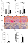
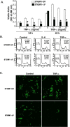
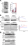
Treatment of multiple myeloma with bortezomib can result in severe adverse effects, necessitating the development of targeted inhibitors of the proteasome. We show that stable expression of a dominant-negative F-box deleted (âF) mutant of the E3 ubiquitin ligase, SCFβ-TrCP/FWD1, in murine 5TGM1 myeloma cells dramatically attenuated their skeletal engraftment and survival when inoculated into immunocompetent C57BL/KaLwRij mice. Similar results were obtained in immunodeficient bg-nu-xid mice, suggesting that the observed effects were independent of host recipient immune status. Bone marrow stroma offered no protection for 5TGM1-âF cells in cocultures treated with tumor necrosis factor (TNF), indicating a cell-autonomous anti-myeloma effect. Levels of p100, IκBα, Mcl-1, ATF4, total and cleaved caspase-3, and phospho-β-catenin were elevated in 5TGM1-âF cells whereas cIAP was down-regulated. TNF also activated caspase-3 and downregulated Bcl-2, correlating with the enhanced susceptibility of 5TGM1-âF cells to apoptosis. Treatment of 5TGM1 tumor-bearing mice with a β-TrCP1/FWD1 inhibitor, pyrrolidine dithiocarbamate (PDTC), significantly reduced tumor burden in bone. PDTC also increased levels of cleaved Mcl-1 and caspase-3 in U266 human myeloma cells, correlating with our murine data and validating the development of specific β-TrCP inhibitors as an alternative therapy to nonspecific proteasome inhibitors for myeloma patients.
Keywords: 5TGM1; FWD1/β-TrCP1; myeloma; plasmacytoma; proteasome.
Conflict of interest statement
There is no conflict of interest.
Figures


Similar articles
-
Associations among beta-TrCP, an E3 ubiquitin ligase receptor, beta-catenin, and NF-kappaB in colorectal cancer.J Natl Cancer Inst. 2004 Aug 4;96(15):1161-70. doi: 10.1093/jnci/djh219. J Natl Cancer Inst. 2004. PMID: 15292388
-
SAG/ROC-SCF beta-TrCP E3 ubiquitin ligase promotes pro-caspase-3 degradation as a mechanism of apoptosis protection.Neoplasia. 2006 Dec;8(12):1042-54. doi: 10.1593/neo.06568. Neoplasia. 2006. PMID: 17217622 Free PMC article.
-
Mathematical modelling suggests a differential impact of β-transducin repeat-containing protein paralogues on Wnt/β-catenin signalling dynamics.FEBS J. 2015 Mar;282(6):1080-96. doi: 10.1111/febs.13204. Epub 2015 Feb 7. FEBS J. 2015. PMID: 25601154
-
Surgical thyroparathyroidectomy prevents progression of 5TGM1 murine multiple myeloma in vivo.J Bone Oncol. 2018 Feb 23;12:19-22. doi: 10.1016/j.jbo.2018.02.005. eCollection 2018 Sep. J Bone Oncol. 2018. PMID: 29556454 Free PMC article. Review.
-
Small molecule and peptide inhibitors of βTrCP and the βTrCP-NRF2 protein-protein interaction.Biochem Soc Trans. 2023 Jun 28;51(3):925-936. doi: 10.1042/BST20220352. Biochem Soc Trans. 2023. PMID: 37293994 Free PMC article. Review.
Cited by
-
β-TrCP1 promotes cell proliferation via TNF-dependent NF-κB activation in diffuse large B cell lymphoma.Cancer Biol Ther. 2020;21(3):241-247. doi: 10.1080/15384047.2019.1683332. Epub 2019 Nov 15. Cancer Biol Ther. 2020. PMID: 31731887 Free PMC article.
-
Genetic determinants of multiple myeloma risk within the Wnt/beta-catenin signaling pathway.Cancer Epidemiol. 2021 Aug;73:101972. doi: 10.1016/j.canep.2021.101972. Epub 2021 Jun 30. Cancer Epidemiol. 2021. PMID: 34216957 Free PMC article.
-
MicroRNA-324-5p suppresses the migration and invasion of MM cells by inhibiting the SCFβ-TrCP E3 ligase.Oncol Lett. 2018 Oct;16(4):5331-5338. doi: 10.3892/ol.2018.9245. Epub 2018 Aug 1. Oncol Lett. 2018. PMID: 30250603 Free PMC article.
-
Decoys Untangle Complicated Redundancy and Reveal Targets of Circadian Clock F-Box Proteins.Plant Physiol. 2018 Jul;177(3):1170-1186. doi: 10.1104/pp.18.00331. Epub 2018 May 23. Plant Physiol. 2018. PMID: 29794020 Free PMC article.
-
Beta-Transducin Repeats-Containing Proteins as an Anticancer Target.Cancers (Basel). 2023 Aug 24;15(17):4248. doi: 10.3390/cancers15174248. Cancers (Basel). 2023. PMID: 37686524 Free PMC article. Review.
References
-
- Glickman MH, Ciechanover A. The ubiquitin-proteasome proteolytic pathway: destruction for the sake of construction. Physiol Rev. 2002;82:373–428. - PubMed
-
- Sahasrabuddhe AA, Elenitoba-Johnson KS. Role of the ubiquitin proteasome system in hematologic malignancies. Immunol Rev. 2015;263:224–239. - PubMed
-
- Dimopoulos MA, Richardson PG, Moreau P, Anderson KC. Current treatment landscape for relapsed and/or refractory multiple myeloma. Nat Rev Clin Oncol. 2014 - PubMed
Publication types
MeSH terms
Substances
Grants and funding
LinkOut - more resources
Full Text Sources
Other Literature Sources
Medical
Research Materials

