

Exosome enrichment of human serum using multiple cycles of centrifugation
- PMID: 26010067
- PMCID: PMC4558199
- DOI: 10.1002/elps.201500131
Exosome enrichment of human serum using multiple cycles of centrifugation
Abstract
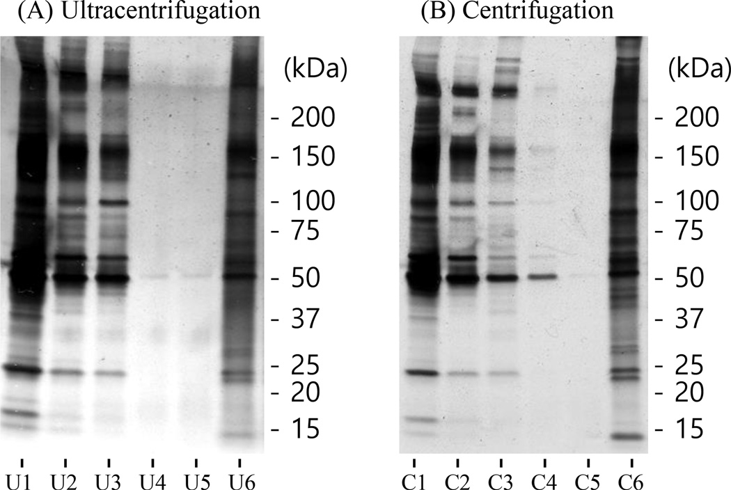
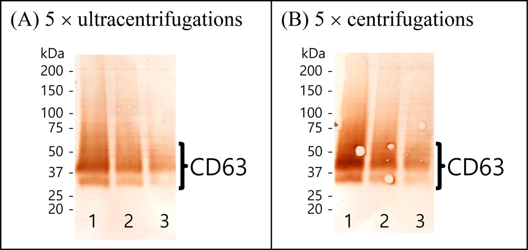
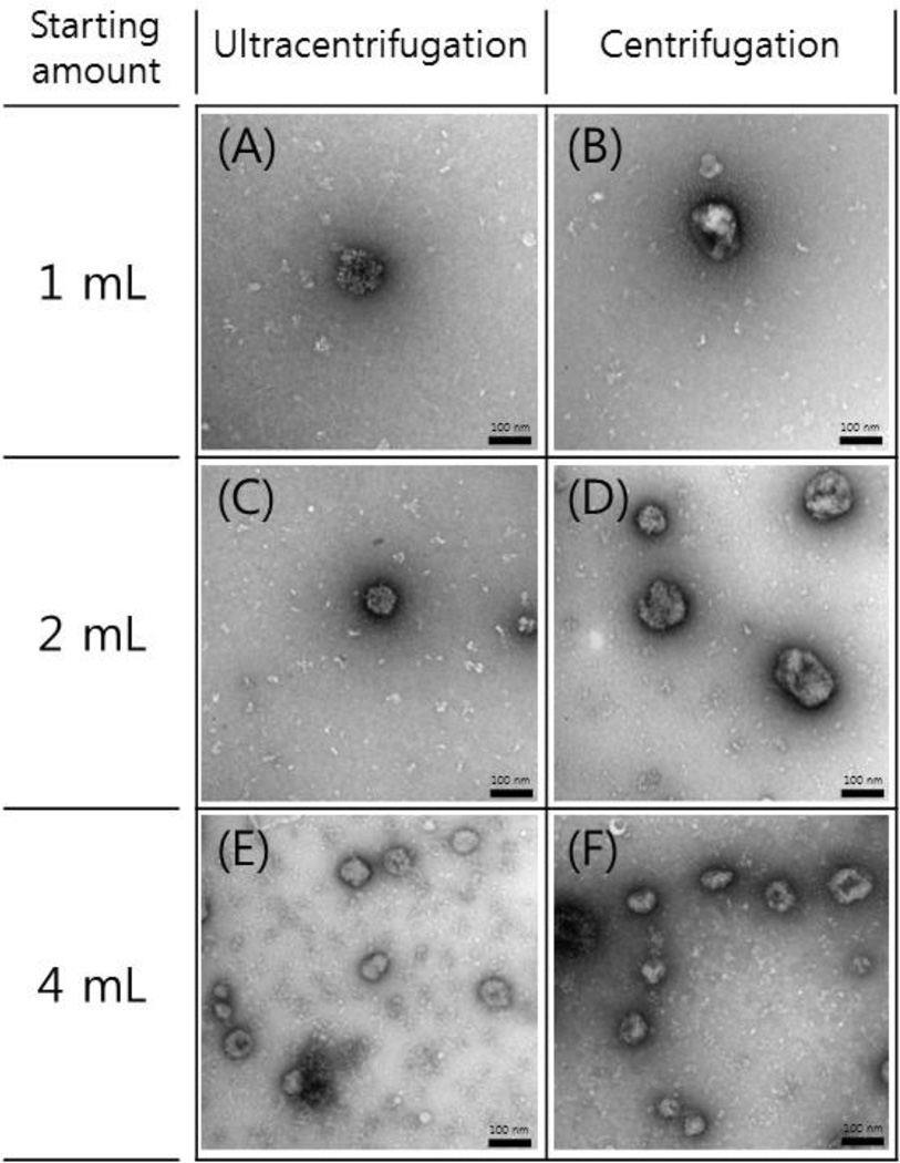
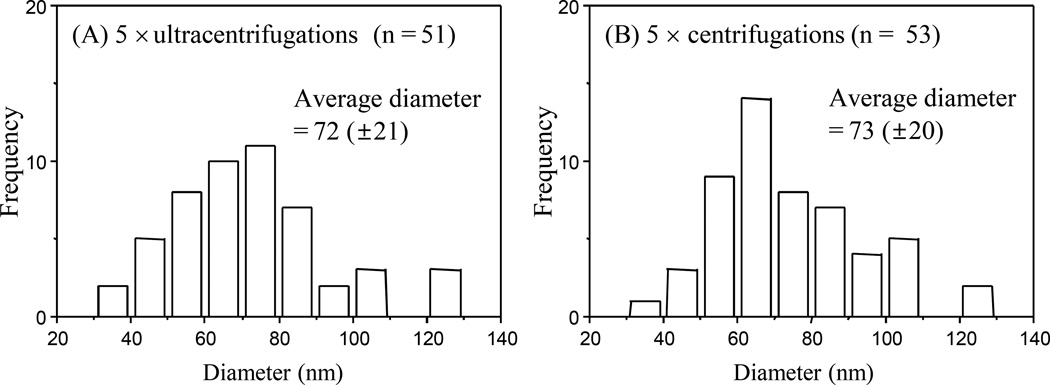
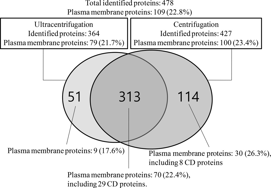
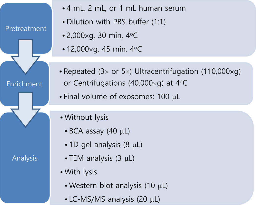
In this work, we compared the use of repeated cycles of centrifugation at conventional speeds for enrichment of exosomes from human serum compared to the use of ultracentrifugation (UC). After removal of cells and cell debris, a speed of 110 000 × g or 40 000 × g was used for the UC or centrifugation enrichment process, respectively. The enriched exosomes were analyzed using the bicinchoninic acid assay, 1D gel separation, transmission electron microscopy, Western blotting, and high-resolution LC-MS/MS analysis. It was found that a five-cycle repetition of UC or centrifugation is necessary for successful removal of nonexosomal proteins in the enrichment of exosomes from human serum. More significantly, 5× centrifugation enrichment was found to provide similar or better performance than 5× UC enrichment in terms of enriched exosome protein amount, Western blot band intensity for detection of CD-63, and numbers of identified exosome-related proteins and cluster of differentiation (CD) proteins. A total of 478 proteins were identified in the LC-MS/MS analyses of exosome proteins obtained from 5× UCs and 5× centrifugations including many important CD membrane proteins. The presence of previously reported exosome-related proteins including key exosome protein markers demonstrates the utility of this method for analysis of proteins in human serum.
Keywords: Centrifugation; Exosomes; Human serum; Mass spectrometry; Ultracentrifugation.
© 2015 WILEY-VCH Verlag GmbH & Co. KGaA, Weinheim.
Conflict of interest statement
The authors have declared no conflict of interest.
Figures
Similar articles
-
Comparison of an Optimized Ultracentrifugation Method versus Size-Exclusion Chromatography for Isolation of Exosomes from Human Serum.J Proteome Res. 2018 Oct 5;17(10):3599-3605. doi: 10.1021/acs.jproteome.8b00479. Epub 2018 Sep 19. J Proteome Res. 2018. PMID: 30192545 Free PMC article.
-
A protocol for exosome isolation and characterization: evaluation of ultracentrifugation, density-gradient separation, and immunoaffinity capture methods.Methods Mol Biol. 2015;1295:179-209. doi: 10.1007/978-1-4939-2550-6_15. Methods Mol Biol. 2015. PMID: 25820723
-
An improvised one-step sucrose cushion ultracentrifugation method for exosome isolation from culture supernatants of mesenchymal stem cells.Stem Cell Res Ther. 2018 Jul 4;9(1):180. doi: 10.1186/s13287-018-0923-0. Stem Cell Res Ther. 2018. PMID: 29973270 Free PMC article.
-
Integrated systems for exosome investigation.Methods. 2015 Oct 1;87:31-45. doi: 10.1016/j.ymeth.2015.04.015. Epub 2015 Apr 24. Methods. 2015. PMID: 25916618 Review.
-
Techniques Associated with Exosome Isolation for Biomarker Development: Liquid Biopsies for Ovarian Cancer Detection.Methods Mol Biol. 2020;2055:181-199. doi: 10.1007/978-1-4939-9773-2_8. Methods Mol Biol. 2020. PMID: 31502152 Review.
Cited by
-
Involvement of serum-derived exosomes of elderly patients with bone loss in failure of bone remodeling via alteration of exosomal bone-related proteins.Aging Cell. 2018 Jun;17(3):e12758. doi: 10.1111/acel.12758. Epub 2018 Mar 30. Aging Cell. 2018. PMID: 29603567 Free PMC article.
-
Exosomes play an important role in the process of psoralen reverse multidrug resistance of breast cancer.J Exp Clin Cancer Res. 2016 Dec 1;35(1):186. doi: 10.1186/s13046-016-0468-y. J Exp Clin Cancer Res. 2016. PMID: 27906043 Free PMC article.
-
A novel method of high-purity extracellular vesicle enrichment from microliter-scale human serum for proteomic analysis.Electrophoresis. 2021 Feb;42(3):245-256. doi: 10.1002/elps.202000223. Electrophoresis. 2021. PMID: 33169421 Free PMC article.
-
Comparison of an Optimized Ultracentrifugation Method versus Size-Exclusion Chromatography for Isolation of Exosomes from Human Serum.J Proteome Res. 2018 Oct 5;17(10):3599-3605. doi: 10.1021/acs.jproteome.8b00479. Epub 2018 Sep 19. J Proteome Res. 2018. PMID: 30192545 Free PMC article.
-
High-Performance Chemical Isotope Labeling Liquid Chromatography Mass Spectrometry for Exosome Metabolomics.Anal Chem. 2018 Jul 17;90(14):8314-8319. doi: 10.1021/acs.analchem.8b01726. Epub 2018 Jul 3. Anal Chem. 2018. PMID: 29920066 Free PMC article.
References
-
- Caby M-P, Lankar D, Vincendeau-Scherrer C, Raposo G, Bonnerot C. Int. Immuno. 2005;17:879–887. - PubMed
-
- Kalra H, Adda CG, Liem M, Ang CS, Mechler A, Simpson RJ, Hulett MD, Mathivanan S. Proteomics. 2013;13:3354–3364. - PubMed
-
- Moon PG, You S, Lee JE, Hwang D, Baek MC. Mass Spectrom. Rev. 2011;30:1185–1202. - PubMed
-
- Moon P-G, Lee J-E, You S, Kim T-K, Cho J-H, Kim I-S, Kwon T-H, Kim C-D, Park S-H, Hwang D, Kim Y-L, Baek M-C. Proteomics. 2011;11:2459–2475. - PubMed
Publication types
MeSH terms
Substances
Grants and funding
LinkOut - more resources
Full Text Sources
Other Literature Sources
Miscellaneous
