Clinical Trial
. 2015 May 26;10(5):e0127934.
doi: 10.1371/journal.pone.0127934. eCollection 2015.

Physiologically Based Pharmacokinetic Modeling Is Essential in 90Y-Labeled Anti-CD66 Radioimmunotherapy

Affiliations
Clinical Trial

Physiologically Based Pharmacokinetic Modeling Is Essential in 90Y-Labeled Anti-CD66 Radioimmunotherapy

Peter Kletting et al. PLoS One. .

Abstract

Introduction: Radioimmunotherapy (RIT) with 90Y-labeled anti-CD66 antibody is used to selectively irradiate the red marrow (RM) before blood stem cell transplantation of acute leukemia patients. To calculate the activity to administer, time-integrated activity coefficients are required. These are estimated prior to therapy using gamma camera and serum measurements after injection of 111In labeled anti-CD66 antibody. Equal pre-therapeutic and therapeutic biodistributions are usually assumed to calculate the coefficients. However, additional measurements during therapy had shown that this assumption had to be abandoned. A physiologically based pharmacokinetic (PBPK) model was developed to allow the prediction of therapeutic time-integrated activity coefficients in eight patients.

Aims: The aims of the study were to demonstrate using a larger patient group 1) the need to perform patient-specific dosimetry in 90Y-labeled anti-CD66 RIT, 2) that pre-therapeutic and therapeutic biodistributions differ, and most importantly 3) that this difference in biodistributions can be accurately predicted using a refined model.

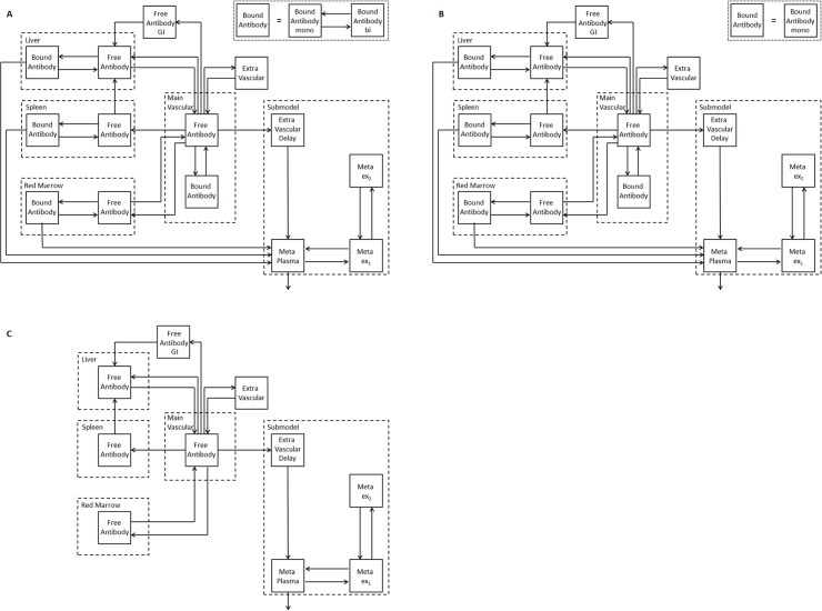
Materials and methods: Two new PBPK models were developed considering fully, half and non-immunoreactive antibodies and constraints for estimating the RM antigen number. Both models were fitted to gamma camera and serum measurements of 27 patients. Akaike weights were used for model averaging. Time-integrated activity coefficients for total body, liver, spleen, RM and serum were calculated. Model-based predictions of the serum biokinetics during therapy were compared to actual measurements.

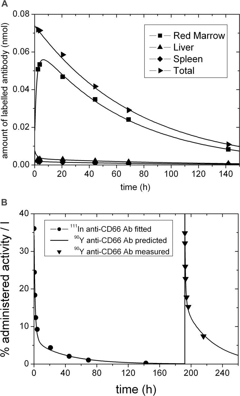
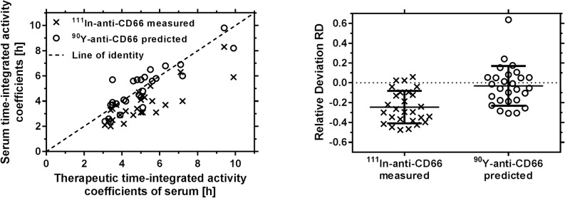
Results: Variability of the RM time-integrated activity coefficients ((37.3 ± 7.5) h) indicates the need for patient-specific dosimetry. The relative differences between pre-therapeutic and therapeutic serum time-activity curves were (-25 ± 16)%. The prediction accuracy of these differences using the refined PBPK models was (-3 ± 20)%.

Conclusion: Individual treatment is needed due to biological differences between patients in RIT with 90Y-labeled anti-CD66 antibody. Differences in pre-therapeutic and therapeutic biokinetics are predominantly caused by different degrees of saturation due to different amounts of administered antibody. These differences could be predicted using the PBPK models.


Conflict of interest statement

Competing Interests: The authors have declared that no competing interests exist.

Figures

Fig 1
Fig 1. PBPK model for radiolabeled anti-CD66 monoclonal antibodies.
Models for (A) fully intact (both antigen-binding sites are active, i.e. bivalent binding of antibody possible), (B) half (one antigen-binding site is active, i.e. monovalent binding) and (C) non-immunoreactive antibody (both antigen-binding sites are inactive, i.e. no binding). Due to the equivalence of both valences of the antibody, the fractions of antibodies in (A), (B) and (C) are determined as follows: With the probability rim of one antibody valence being immunoreactive, the fractions of fully, half or non-immunoreactive antibody injected in (A), (B) and (C) are rim2,2rim(1rim), and (1 − r im)2, respectively. The model consists of two equal subsystems describing the biodistribution of the labeled and unlabeled antibodies (this is true for A, B and C). The labeled and unlabeled species are competing for binding to free antigens (only A and B). The subsystems are additionally connected via physical decay, i.e. when the radiolabel decays the molecule enters the corresponding unlabeled compartment. The corresponding model equations are provided in supplement S1 Text. Radiolabeled and unlabeled antibodies are intravenously injected (main vascular compartment). The antibodies are distributed via blood flow to the main CD66 antigen expression sites. The discontinuous capillary structure of the liver, spleen and the red marrow allows the modeling of the vascular and interstitial space as one compartment. The degradation rate of bound antibody is assumed to be the same in all organs. The submodel for degraded antibody is adopted from Houston et al. [3, 28]. GI = gastrointestinal tract; Meta = metabolites in plasma; ex1, ex2 = extravascular metabolites; mono = monovalent and bi = bivalent binding.
Fig 2
Fig 2. Typical biokinetic data, fit and prediction.
Biokinetic data and the pertaining fitted curves using model 2 (solid lines) for labeled anti-CD66 antibodies (A) in red marrow, liver, spleen and whole body and (B) in serum. The solid line for times larger than 190 h post injection depicts the excellent prediction for the therapeutic time-activity curve based on the fitted parameters of model 2 using pre-therapeutic data only. Note that for this patient no 48 h measurement was obtained. The corresponding red marrow kinetics for all patients are presented in supplement S2 Fig.
Fig 3
Fig 3. Serum time-integrated activity coefficients.
(Left) Measured pre-therapeutic (111In) and predicted therapeutic (90Y) serum measurements versus actual therapeutic time-integrated activity coefficients of all patients. The application of the PBPK model allows for the prediction of therapeutic serum time-activity curves and removes the systematic offset. (Right) Relative deviation of serum time-integrated activity coefficients for all patients (scatterplots with mean and standard deviations).

References

    1. Sharkey RM, Goldenberg DM. Cancer radioimmunotherapy. Immunotherapy. 2011;3(3):349–70. 10.2217/imt.10.114 PubMed - DOI - PMC - PubMed
    1. Abutalib S.A., Tallman M.S. Monoclonal Antibodies for the Treatment of Acute Myeloid Leukemia. Curr Pharm Biotechnol. 2006;7:343–69. - PubMed
    1. Kletting P, Kull T, Bunjes D, Mahren B, Luster M, Reske SN, et al. Radioimmunotherapy with anti-CD66 antibody: Improving the biodistribution using a physiologically based pharmacokinetic model. J Nucl Med. 2010;51(3):484–91. 10.2967/jnumed.109.067546 - DOI - PubMed
    1. Schulz A, Glatting G, Hoenig M, Schuetz C, Gatz S, Grewendorf S, et al. Radioimmunotherapy-based conditioning for hematopoietic cell transplantation in children with malignant and nonmalignant diseases. Blood. 2011;117(17):4642–50. 10.1182/blood-2010-06-284349 PubMed . - DOI - PubMed
    1. Ringhoffer M, Blumstein N, Neumaier B, Glatting G, von Harsdorf S, Buchmann I, et al. 188Re- or 90Y-labelled anti-CD66 antibody as part of a dose-reduced conditioning regimen for patients with acute leukaemia or myelodysplastic syndrome over the age of 55: results of a phase I-II study. Br J Haematol. 2005;130(4):604–13. - PubMed

Publication types