

Characterization of the Copper(II) Binding Sites in Human Carbonic Anhydrase II
- PMID: 26010488
- PMCID: PMC4482258
- DOI: 10.1021/acs.inorgchem.5b00057
Characterization of the Copper(II) Binding Sites in Human Carbonic Anhydrase II
Abstract
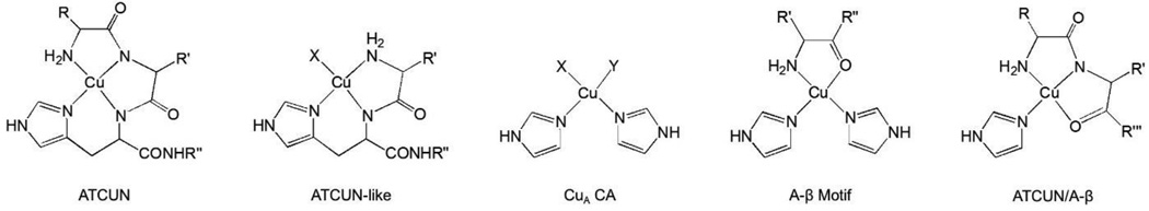
Human carbonic anhydrase (CA) is a well-studied, robust, mononuclear Zn-containing metalloprotein that serves as an excellent biological ligand system to study the thermodynamics associated with metal ion coordination chemistry in aqueous solution. The apo form of human carbonic anhydrase II (CA) binds 2 equiv of copper(II) with high affinity. The Cu(2+) ions bind independently forming two noncoupled type II copper centers in CA (CuA and CuB). However, the location and coordination mode of the CuA site in solution is unclear, compared to the CuB site that has been well-characterized. Using paramagnetic NMR techniques and X-ray absorption spectroscopy we identified an N-terminal Cu(2+) binding location and collected information on the coordination mode of the CuA site in CA, which is consistent with a four- to five-coordinate N-terminal Cu(2+) binding site reminiscent to a number of N-terminal copper(II) binding sites including the copper(II)-amino terminal Cu(2+) and Ni(2+) and copper(II)-β-amyloid complexes. Additionally, we report a more detailed analysis of the thermodynamics associated with copper(II) binding to CA. Although we are still unable to fully deconvolute Cu(2+) binding data to the high-affinity CuA site, we derived pH- and buffer-independent values for the thermodynamics parameters K and ΔH associated with Cu(2+) binding to the CuB site of CA to be 2 × 10(9) and -17.4 kcal/mol, respectively.
Figures


Similar articles
-
Building reactive copper centers in human carbonic anhydrase II.J Biol Inorg Chem. 2013 Aug;18(6):595-8. doi: 10.1007/s00775-013-1009-1. Epub 2013 Jun 7. J Biol Inorg Chem. 2013. PMID: 23744511 Free PMC article.
-
Revisiting zinc coordination in human carbonic anhydrase II.Inorg Chem. 2012 Oct 15;51(20):11098-105. doi: 10.1021/ic301645j. Epub 2012 Oct 3. Inorg Chem. 2012. PMID: 23030313 Free PMC article.
-
Metal Ion Binding Induces Local Protein Unfolding and Destabilizes Human Carbonic Anhydrase II.Inorg Chem. 2022 Jan 24;61(3):1249-1253. doi: 10.1021/acs.inorgchem.1c03271. Epub 2022 Jan 6. Inorg Chem. 2022. PMID: 34989562 Free PMC article.
-
Structurally distinct active sites in the copper(II)-substituted aminopeptidases from Aeromonas proteolytica and Escherichia coli.J Am Chem Soc. 2002 Nov 6;124(44):13025-34. doi: 10.1021/ja026341p. J Am Chem Soc. 2002. PMID: 12405829 Free PMC article.
-
Carbonic anhydrase II-based metal ion sensing: Advances and new perspectives.Biochim Biophys Acta. 2010 Feb;1804(2):393-403. doi: 10.1016/j.bbapap.2009.09.031. Epub 2009 Oct 8. Biochim Biophys Acta. 2010. PMID: 19818877 Free PMC article. Review.
Cited by
-
Global stability of an α-ketoglutarate-dependent dioxygenase (TauD) and its related complexes.Biochim Biophys Acta Gen Subj. 2017 May;1861(5 Pt A):987-994. doi: 10.1016/j.bbagen.2017.02.018. Epub 2017 Feb 15. Biochim Biophys Acta Gen Subj. 2017. PMID: 28214548 Free PMC article.
-
Energetics and dynamics of the proton shuttle of carbonic anhydrase II.Cell Mol Life Sci. 2023 Sep 9;80(10):286. doi: 10.1007/s00018-023-04936-z. Cell Mol Life Sci. 2023. PMID: 37688664 Free PMC article.
-
Stress-Related Changes in the Expression and Activity of Plant Carbonic Anhydrases.Planta. 2021 Feb 3;253(2):58. doi: 10.1007/s00425-020-03553-5. Planta. 2021. PMID: 33532871 Review.
-
The Complex Relationship between Metals and Carbonic Anhydrase: New Insights and Perspectives.Int J Mol Sci. 2016 Jan 19;17(1):127. doi: 10.3390/ijms17010127. Int J Mol Sci. 2016. PMID: 26797606 Free PMC article. Review.
-
Long-range paramagnetic NMR data can provide a closer look on metal coordination in metalloproteins.J Biol Inorg Chem. 2018 Jan;23(1):71-80. doi: 10.1007/s00775-017-1511-y. Epub 2017 Dec 7. J Biol Inorg Chem. 2018. PMID: 29218635
References
Publication types
MeSH terms
Substances
Grants and funding
LinkOut - more resources
Full Text Sources
Other Literature Sources
