. 2015 May 26;10(5):e0127421.
doi: 10.1371/journal.pone.0127421. eCollection 2015.

Potential interactions of calcium-sensitive reagents with zinc ion in different cultured cells

Affiliations

Potential interactions of calcium-sensitive reagents with zinc ion in different cultured cells

Koichi Fujikawa et al. PLoS One. .

Abstract

Background: Several chemicals have been widely used to evaluate the involvement of free Ca(2+) in mechanisms underlying a variety of biological responses for decades. Here, we report high reactivity to zinc of well-known Ca(2+)-sensitive reagents in diverse cultured cells.

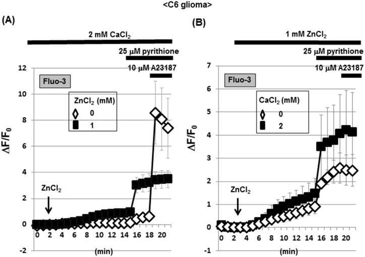
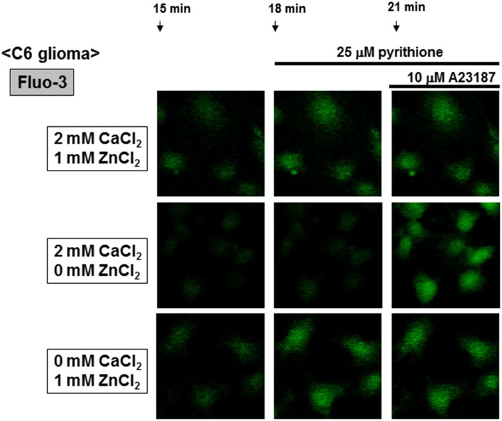
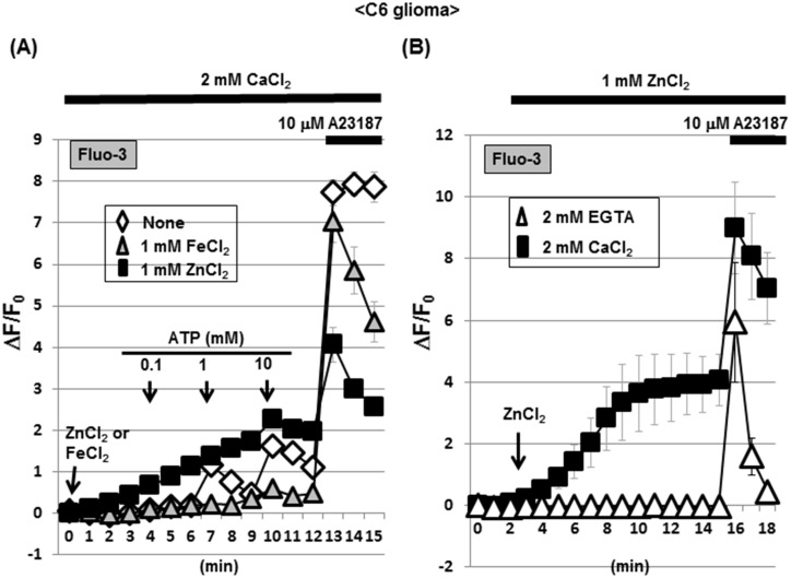
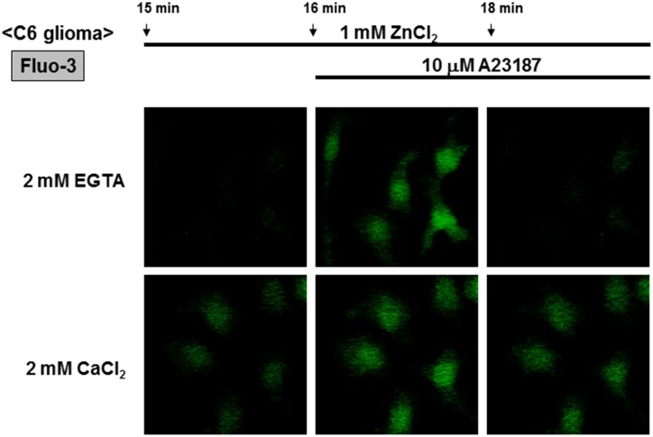
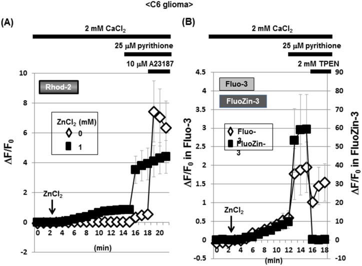
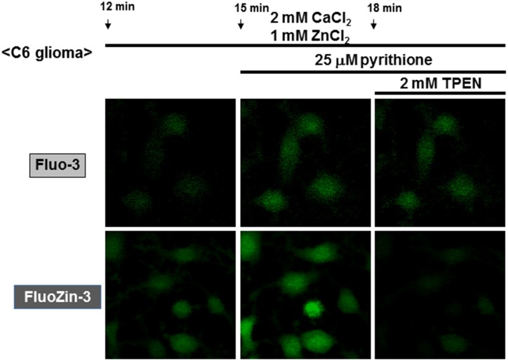
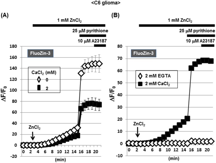
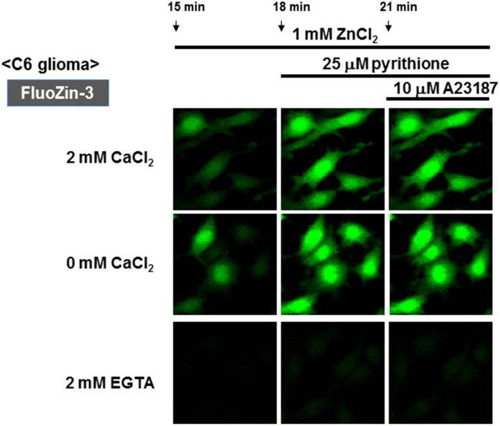
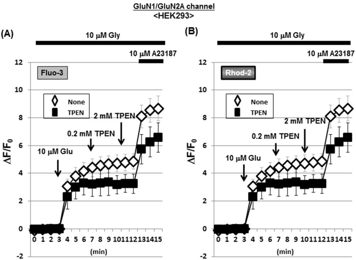
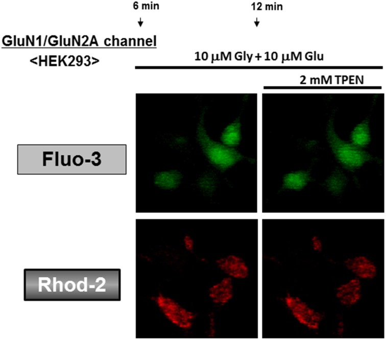
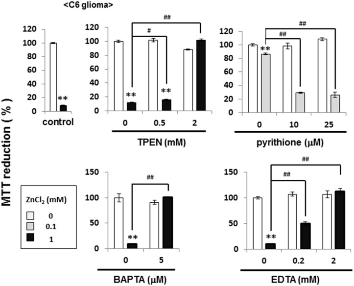
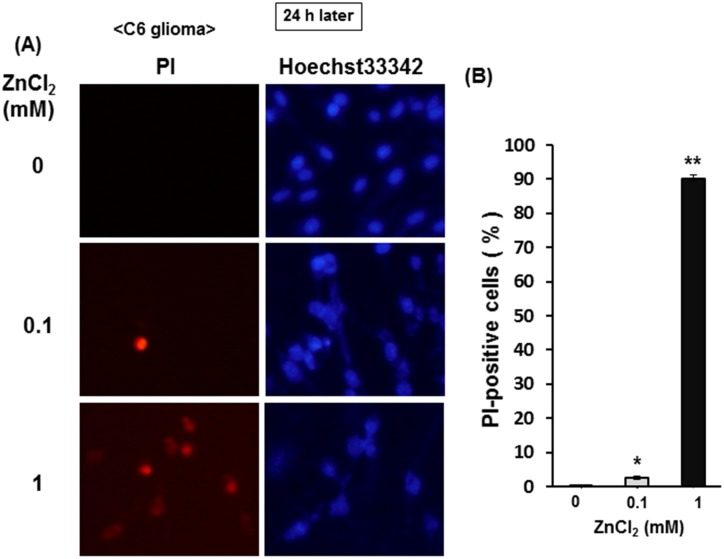
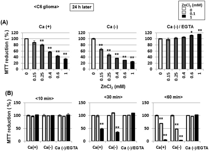
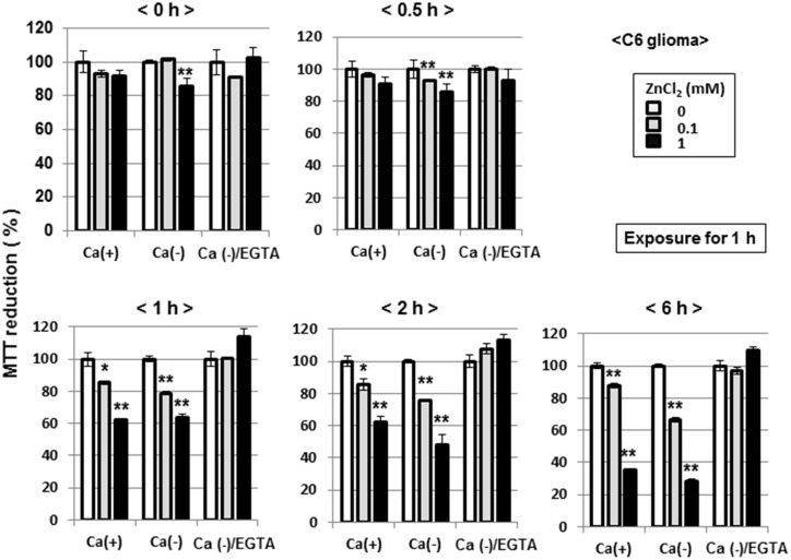
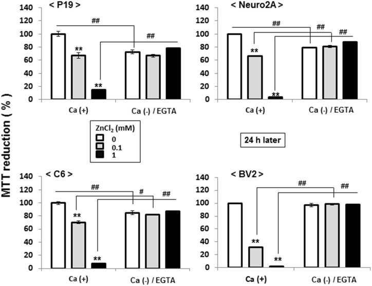
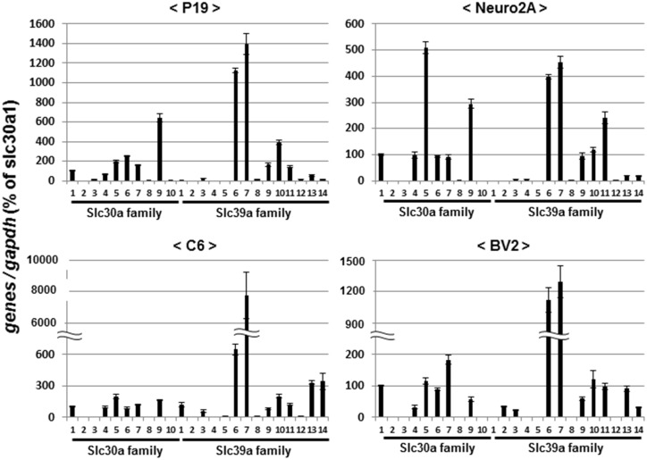
Methodology/principal findings: In rat astrocytic C6 glioma cells loaded with the fluorescent Ca(2+) dye Fluo-3, the addition of ZnCl2 gradually increased the fluorescence intensity in a manner sensitive to the Ca(2+) chelator EGTA irrespective of added CaCl2. The addition of the Ca(2+) ionophore A23187 drastically increased Fluo-3 fluorescence in the absence of ZnCl2, while the addition of the Zn(2+) ionophore pyrithione rapidly and additionally increased the fluorescence in the presence of ZnCl2, but not in its absence. In cells loaded with the zinc dye FluoZin-3 along with Fluo-3, a similarly gradual increase was seen in the fluorescence of Fluo-3, but not of FluoZin-3, in the presence of both CaCl2 and ZnCl2. Further addition of pyrithione drastically increased the fluorescence intensity of both dyes, while the addition of the Zn(2+) chelator N,N,N',N'-tetrakis(2-pyridylmethyl)ethane-1,2-diamine (TPEN) rapidly and drastically decreased FluoZin-3 fluorescence. In cells loaded with FluoZin-3 alone, the addition of ZnCl2 induced a gradual increase in the fluorescence in a fashion independent of added CaCl2 but sensitive to EGTA. Significant inhibition was found in the vitality to reduce 3-(4,5-dimethyl-2-thiazolyl)-2,5-diphenyl-2H-tetrazolium bromide in a manner sensitive to TPEN, EDTA and BAPTA in C6 glioma cells exposed to ZnCl2, with pyrithione accelerating the inhibition. Similar inhibition occurred in an EGTA-sensitive fashion after brief exposure to ZnCl2 in pluripotent P19 cells, neuronal Neuro2A cells and microglial BV2 cells, which all expressed mRNA for particular zinc transporters.

Conclusions/significance: Taken together, comprehensive analysis is absolutely required for the demonstration of a variety of physiological and pathological responses mediated by Ca(2+) in diverse cells enriched of Zn(2+).


Conflict of interest statement

Competing Interests: The authors have declared that no competing interests exist.

Figures

Fig 1
Fig 1. Effects of ZnCl2 on Fluo-3 fluorescence in C6 glioma cells.
(A) C6 glioma cells were cultured for 24 h, followed by loading of Fluo-3 in the presence of CaCl2 and subsequent determination of the fluorescence intensity in either the presence or absence of ZnCl2 every 1 min for 21 min. (B) Cells were loaded with Fluo-3 in either the presence or absence of CaCl2, followed by determination of the fluorescence intensity in the presence of ZnCl2 every 1 min. Values are the mean±S.E. of the rate of fluorescence change in 3 different experiments.
Fig 2
Fig 2. Micrographic pictures of Fluo-3 fluorescence in C6 glioma cells exposed to CaCl2 and ZnCl2 in the presence of A23187 and pyrithione.
Typical pictures are shown here.
Fig 3
Fig 3. A selective increase by ZnCl2 in Fluo-3 fluorescence in C6 glioma cells.
(A) Cells were loaded with Fluo-3 in the presence of CaCl2, followed by determination of the fluorescence intensity in either the presence or absence of ZnCl2 and FeCl2 every 1 min. Cells were exposed to ATP at different concentrations during the determination of fluorescence. (B) Cells were loaded with Fluo-3 in the presence of either EGTA or CaCl2, followed by determination of the fluorescence intensity in the presence of ZnCl2 every 1 min. Values are the mean±S.E. of the rate of fluorescence change in 3 different experiments.
Fig 4
Fig 4. Micrographic pictures of Fluo-3 fluorescence in C6 glioma cells exposed to CaCl2 and ZnCl2 in the presence of A23187 and EGTA.
Typical pictures are shown here.
Fig 5
Fig 5. Possible interaction of Ca2+-sensitive dyes with Zn2+ in C6 glioma cells.
(A) C6 glioma cells were loaded with Rhod-2 in the presence of CaCl2, followed by determination of the fluorescence intensity in either the presence or absence of ZnCl2 every 1 min. (B) Cells were loaded with either Fluo-3 or FluoZin-3 in the presence of CaCl2, followed by determination of the fluorescence intensity in the presence of ZnCl2 every 1 min. Values are the mean±S.E. of the rate of fluorescence change in 3 different experiments.
Fig 6
Fig 6. Micrographic pictures of Rhod-2 fluorescence in C6 glioma cells exposed to CaCl2 and ZnCl2 in the presence of pyrithione.
Typical pictures are shown here.
Fig 7
Fig 7. Micrographic pictures of both Fluo-3 and FluoZin-3 fluorescence in C6 glioma cells exposed to CaCl2 and ZnCl2 in the presence of pyrithione and TPEN.
Typical pictures are shown here.
Fig 8
Fig 8. Effects of ZnCl2 on FluoZin-3 fluorescence in C6 glioma cells.
(A) C6 glioma cells were loaded with FluoZin-3 in either the presence or absence of CaCl2, followed by determination of the fluorescence intensity in the presence of ZnCl2 every 1 min. (B) Cells were loaded with FluoZin-3 in the presence of either EGTA or CaCl2, followed by determination of the fluorescence intensity in the presence of ZnCl2 every 1 min. Values are the mean±S.E. of the rate of fluorescence change in 3 different experiments.
Fig 9
Fig 9. Micrographic pictures of FluoZin-3 fluorescence in C6 glioma cells exposed to CaCl2 and ZnCl2 in the presence of A23187 and pyrithione.
Typical pictures are shown here.
Fig 10
Fig 10. Effects of TPEN on Fluo-3 and Rhod-2 fluorescence in HEK293 cells with acquired NMDAR channels.
HEK293 cells were transfected with expression vectors of GluNR1 and GluNR2A, followed by further culture for an additional 24 h and subsequent loading of either (A) Fluo-3 or (B) Rhod-2 in the presence of Gly. Cells were then exposed to Glu in either the presence or absence of TPEN during the determination of each fluorescence intensity every 1 min. Values are the mean±S.E. of the rate of fluorescence change in 3 different experiments.
Fig 11
Fig 11. Micrographic pictures of both Fluo-3 and FluoZin-3 fluorescence in HEK293 cells with artificial NMDAR.
Typical pictures are shown here.
Fig 12
Fig 12. Effects of ZnCl2 on MTT reducing activity in C6 glioma cells.
Cells were exposed to ZnCl2 at different concentrations in either the presence or absence of TPEN, pyrithione, BAPTA and EDTA for 1 h, followed by culture for an additional 6 h and subsequent determination of MTT reducing activity. Values are the mean±S.E. of percentages over the maximal activity detected in cells not exposed to any test chemicals in 3 different experiments. *P<0.05, **P<0.01, significantly different from the control value in cells not exposed to ZnCl2. #P<0.05, #P<0.01, significantly different from the value in cells exposed to ZnCL2 at each concentration.
Fig 13
Fig 13. Effects of ZnCl2 on PI and Hoechst33342 staining in C6 glioma cells.
Cells were exposed to ZnCl2 at concentrations of 0.1 to 1 mM in the presence of CaCl2 for 1 h, followed by culture for an additional 24 h and subsequent double staining with PI and Hoechst33342 for nuclear DNA. Typical micrographs are shown in the panel (A), while in the panel (B) value are the mean±S.E. of percentages of PI-positive cells over Hoechst33342-positive cells in 3 independent experiments. *P<0.05, **P<0.01, significantly different from the control value in cells not exposed to ZnCl2.
Fig 14
Fig 14. Effects of EGTA on ZnCl2-induced inhibition of MTT reducing activity in C6 glioma cells.
(A) Cells were exposed to ZnCl2 at concentrations of 0.15 to 1 mM in either the presence or absence of CaCl2 and EGTA for 1 h, followed by culture for an additional 24 h and subsequent determination of MTT reducing activity. (B) Cells were also exposed to ZnCl2 at 0.1 or 1 mM in either the presence or absence of CaCl2 and EGTA for different periods from 10 to 60 min, followed by culture for an additional 24 h and subsequent determination of MTT reducing activity. Values are the mean±S.E. of percentages over the maximal activity detected in cells not exposed to any test chemicals in 3 different experiments. *P<0.05, **P<0.01, significantly different from the control value in cells not exposed to ZnCl2.
Fig 15
Fig 15. Effects of culture periods on ZnCl2-induced inhibition of MTT reducing activity in C6 glioma cells.
Cells were exposed to ZnCl2 at 0.1 or 1 mM in either the presence or absence of CaCl2 and EGTA for 1 h, followed by culture for an additional periods from 0.5 to 6 h and subsequent determination of MTT reducing activity. Values are the mean±S.E. of percentages over the maximal activity detected in cells not exposed to any test chemicals in 3 different experiments. *P<0.05, **P<0.01, significantly different from the control value in cells not exposed to ZnCl2.
Fig 16
Fig 16. Effects of ZnCl2 on MTT reducing activity in different cell lines.
Cells were exposed to ZnCl2 at 0.1 or 1 mM in either the presence or absence of CaCl2 and EGTA for 1 h, followed by culture for an additional 24 h and subsequent determination of MTT reducing activity. Values are the mean±S.E. of percentages over the maximal activity detected in cells not exposed to any test chemicals in 3 different experiments. *P<0.05, **P<0.01, significantly different from the control value in cells not exposed to ZnCl2. #P<0.05, #P<0.01, significantly different from the value in cells exposed to ZnCL2 at each concentration.
Fig 17
Fig 17. Expression profiles of Zn2+ transporters in different cell lines.
Cells were cultured under respective appropriate conditions, followed by extraction of total RNA and subsequent determination of mRNA expression on qPCR. Values are the mean±S.E. of percentages over the expression of Slc30a1 in 3 different experiments.

Similar articles

Cited by

References

    1. Scheetz AJ, Constantine-Paton M (1994) Modulation of NMDA receptor function: implications for vertebrate neural development. FASEB J. 8: 745–752. - PubMed
    1. Collingridge GL, Bliss TV (1995) Memories of NMDA receptors and LTP. Trends Neurosci 18: 54–56. - PubMed
    1. Choi DW (1988) Calcium-mediated neurotoxicity: relationship to specific channel types and role in ischemic damage. Trends Neurosci 11: 465–469. - PubMed
    1. Sattler R, Tymianski M (2000) Molecular mechanisms of calcium-dependent excitotoxicity. J Mol Med 78: 3–13. - PubMed
    1. MacDermott AB, Mayer ML, Westbrook GL, Smith SJ, Barker JL (1986) NMDA-receptor activation increases cytoplasmic calcium concentration in cultured spinal cord neurones. Nature 321, 519–522. - PubMed

Publication types

MeSH terms

LinkOut - more resources