. 2015 Dec 15;523(18):2714-37.
doi: 10.1002/cne.23812. Epub 2015 Aug 18.

Projections from the subparaventricular zone define four channels of output from the circadian timing system

Affiliations

Projections from the subparaventricular zone define four channels of output from the circadian timing system

Nina Vujovic et al. J Comp Neurol. .

Abstract

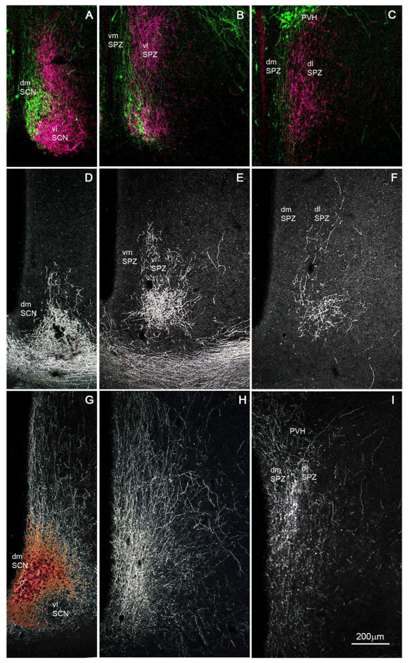
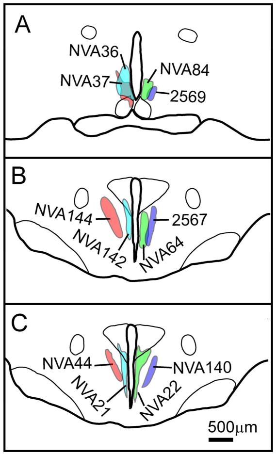
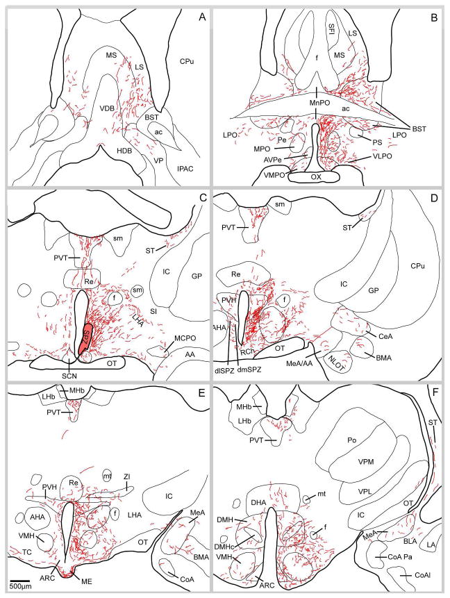
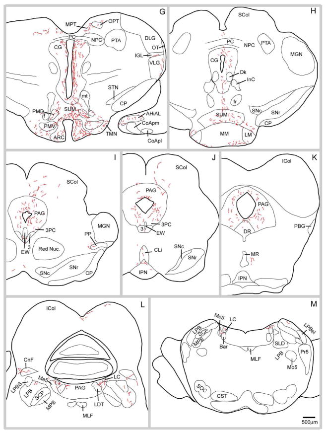
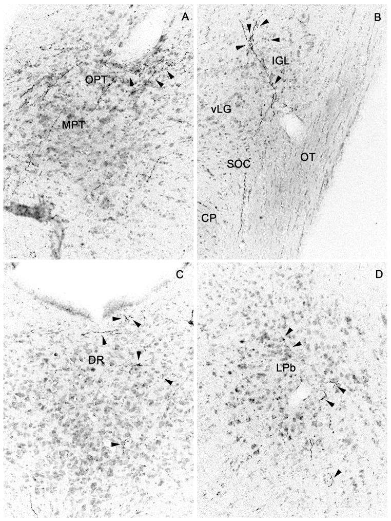
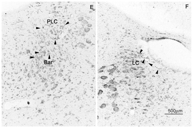
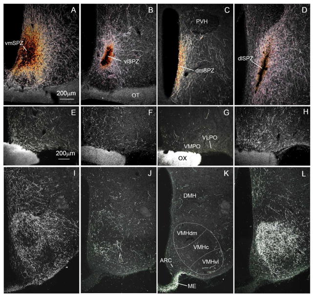
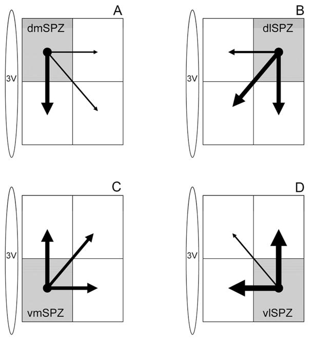
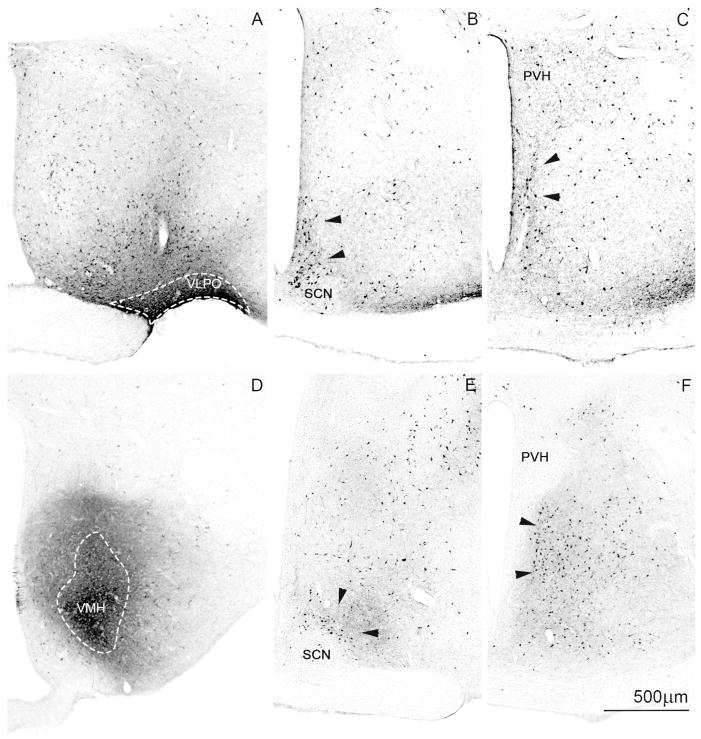
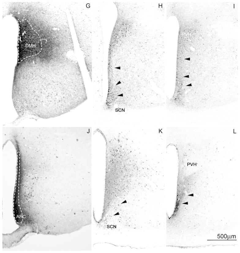
The subparaventricular zone of the hypothalamus (SPZ) is the main efferent target of neural projections from the suprachiasmatic nucleus (SCN) and an important relay for the circadian timing system. Although the SPZ is fairly homogeneous cytoarchitecturally and neurochemically, it has been divided into distinct functional and connectional subdivisions. The dorsal subdivision of the SPZ (dSPZ) plays an important role in relaying signals from the SCN controlling body temperature rhythms, while the ventral subdivision (vSPZ) is critical for rhythms of sleep and locomotor activity (Lu et al. [] J Neurosci 21:4864-4874). On the other hand, the medial part of the SPZ receives input mainly from the dorsomedial SCN, whereas the lateral SPZ receives input from the ventrolateral SCN and the retinohypothalamic tract (Leak and Moore [] J Comp Neurol 433:312-334). We therefore investigated whether there are corresponding differences in efferent outputs from these four quadrants of the SPZ (dorsolateral, ventrolateral, dorsomedial, and ventromedial) by a combination of anterograde and retrograde tracing. We found that, while all four subdivisions of the SPZ share a similar backbone of major projection pathways to the septal region, thalamus, hypothalamus, and brainstem, each segment of the SPZ has a specific set of targets where its projections dominate. Furthermore, we observed intra-SPZ projections of varying densities between the four subdivisions. Taken together, this pattern of organization suggests that the circadian timing system may have several parallel neural outflow pathways that provide a road map for understanding how they subserve different functions.

Keywords: circadian timing; neural projections; subparaventricular zone.


Conflict of interest statement

Conflict of interest

The authors have no conflicts of interest to disclose.

Figures

Figure 1
Figure 1
A series of photomicrographs to illustrate topographic specificity of afferent inputs to the subparaventricular zone. In each row, the photograph at the left (A, D, G) is from the level of the SCN, the one in the middle (B, E, H) is from the level of the rostral/ventral SPZ, and the one on the right (C, F, I) is from the level of the caudal/dorsal SPZ. The first row across (A, B, C) shows afferent from neurons in the SCN expressing arginine vasopressin (green) vs. vasoactive intestinal polypeptide immunoreactivity (magenta). The second row (D, E, F) shows projections from retinal ganglion cells transduced with AAV-GFP (with immunohistochemical enhancement of GFP signal), and the third row (G, H, I) shows inputs from dmSCN neurons transduced with AAV-GFP (with immunohistochemical enhancement of GFP signal).
Figure 2
Figure 2
Camera lucida plots illustrating placement of SPZ injection sites in cases used for comparative analysis across the four SPZ subdivisions at (A) the level of the rostral/ventral SPZ and (B, C) the level of the caudal/dorsal SPZ.
Figure 3
Figure 3
A series of camera lucida drawings illustrating the pattern of axonal labeling in case NV36. Note the presence of fibers in the dorsal raphe, lateral parabrachial nucleus, pre-coeruleus region, locus coeruleus and Barrington’s nucleus.
Figure 3
Figure 3
A series of camera lucida drawings illustrating the pattern of axonal labeling in case NV36. Note the presence of fibers in the dorsal raphe, lateral parabrachial nucleus, pre-coeruleus region, locus coeruleus and Barrington’s nucleus.
Figure 4
Figure 4
Brightfield photomicrographs showing distal SPZ projections (from injection case NVA36; cf. drawings in Fig. 3) to (A) the olivary pretectal nucleus, (B) intergeniculate leaflet, (C) dorsal raphe nucleus, (D) lateral parabrachial nucleus, (E) pre-coeruleus area and Barrington’s nucleus, and (F) locus coeruleus; black arrowheads indicate axons bearing varicosities or terminal boutons.
Figure 4
Figure 4
Brightfield photomicrographs showing distal SPZ projections (from injection case NVA36; cf. drawings in Fig. 3) to (A) the olivary pretectal nucleus, (B) intergeniculate leaflet, (C) dorsal raphe nucleus, (D) lateral parabrachial nucleus, (E) pre-coeruleus area and Barrington’s nucleus, and (F) locus coeruleus; black arrowheads indicate axons bearing varicosities or terminal boutons.
Figure 5
Figure 5
Darkfield photomicrographs showing (A–D) injection sites (scale bar in A, 200μm) and anterogradely labeled fibers and terminals at the level of the preoptic area (E–H) and ventromedial nucleus of the hypothalamus (I–L; scale bar in E, 200μm). Panels A, E, and I show projections from the vmSPZ (case NVA84); B, F, and J show innervation from the vlSPZ (case 2569); C, G, and K show projections from the dmSPZ (case NVA21); and D, H, and L illustrate output from the dlSPZ (case 2567).
Figure 6
Figure 6
Schematic diagram illustrating estimated density of intra-SPZ projections based on anterograde tracing from the (A) dorsomedial, (B) dorsolateral, (C) ventromedial, and (D) ventrolateral SPZ. The thickness of each arrow is roughly proportional to the density of the projection.
Figure 7
Figure 7
Brightfield photomicrographs showing injection sites (A,D,G,J) and retrogradely labeled cells at the level of the vSPZ (B,E,H,K) and dSPZ (C,F,H,L) following CTb injection into the (A–C) ventrolateral preoptic nucleus (case R2059), (D–F) ventromedial hypothalamic nucleus (case NVA151), (G–I) dorsomedial hypothalamic nucleus (case 1927), and (J–L) arcuate nucleus (case NVA150). White dotted lines designate the CTb injection core and black arrowheads designate clusters of retrogradely labeled cells in the SPZ.
Figure 7
Figure 7
Brightfield photomicrographs showing injection sites (A,D,G,J) and retrogradely labeled cells at the level of the vSPZ (B,E,H,K) and dSPZ (C,F,H,L) following CTb injection into the (A–C) ventrolateral preoptic nucleus (case R2059), (D–F) ventromedial hypothalamic nucleus (case NVA151), (G–I) dorsomedial hypothalamic nucleus (case 1927), and (J–L) arcuate nucleus (case NVA150). White dotted lines designate the CTb injection core and black arrowheads designate clusters of retrogradely labeled cells in the SPZ.

References

    1. Aton SJ, Herzog ED. Come together, right…now: synchronization of rhythms in a mammalian circadian clock. Neuron. 2005;48(4):531–534. - PMC - PubMed
    1. Belenky MA, Yarom Y, Pickard GE. Heterogeneous expression of gamma-aminobutyric acid and gamma-aminobutyric acid-associated receptors and transporters in the rat suprachiasmatic nucleus. The Journal of comparative neurology. 2008;506(4):708–732. - PubMed
    1. Bruinstroop E, Cano G, Vanderhorst VG, Cavalcante JC, Wirth J, Sena-Esteves M, Saper CB. Spinal projections of the A5, A6 (locus coeruleus), and A7 noradrenergic cell groups in rats. The Journal of comparative neurology 2011 - PMC - PubMed
    1. Cambras T, Weller JR, Angles-Pujoras M, Lee ML, Christopher A, Diez-Noguera A, Krueger JM, de la Iglesia HO. Circadian desynchronization of core body temperature and sleep stages in the rat. Proceedings of the National Academy of Sciences of the United States of America. 2007;104(18):7634–7639. - PMC - PubMed
    1. Canteras NS, Ribeiro-Barbosa ER, Goto M, Cipolla-Neto J, Swanson LW. The retinohypothalamic tract: comparison of axonal projection patterns from four major targets. Brain research reviews. 2011;65(2):150–183. - PubMed

Publication types