. 2015 May 26;10(5):e0125269.
doi: 10.1371/journal.pone.0125269. eCollection 2015.

Tumor treating fields perturb the localization of septins and cause aberrant mitotic exit

Affiliations

Tumor treating fields perturb the localization of septins and cause aberrant mitotic exit

Nidhi Gera et al. PLoS One. .

Abstract

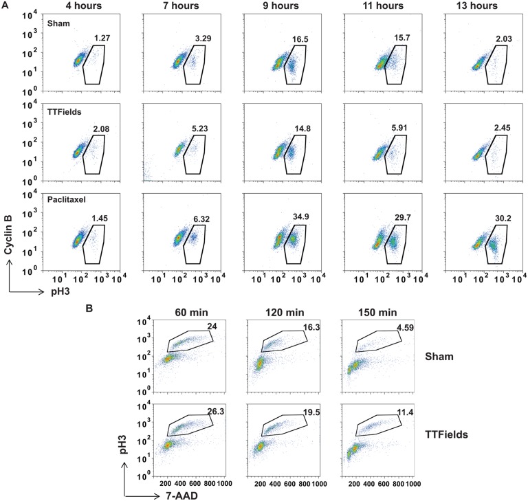
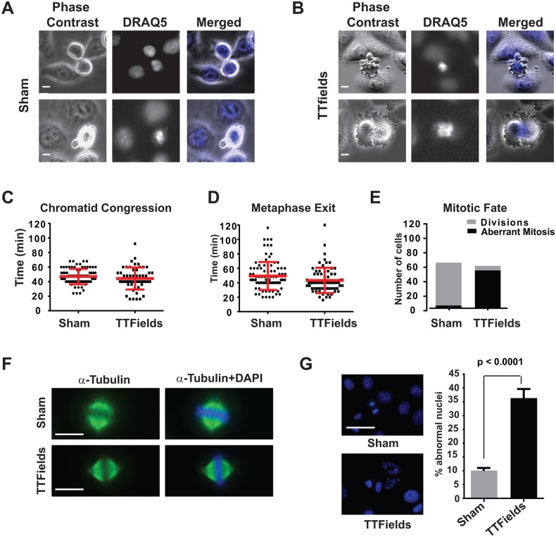
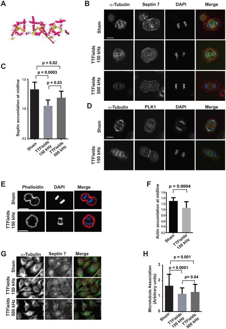
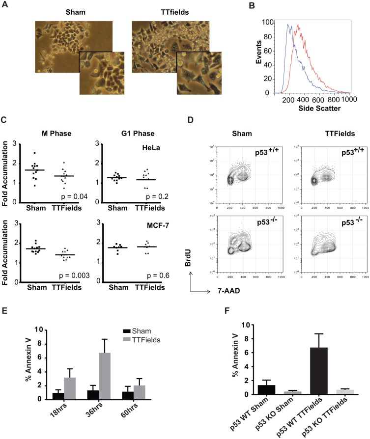
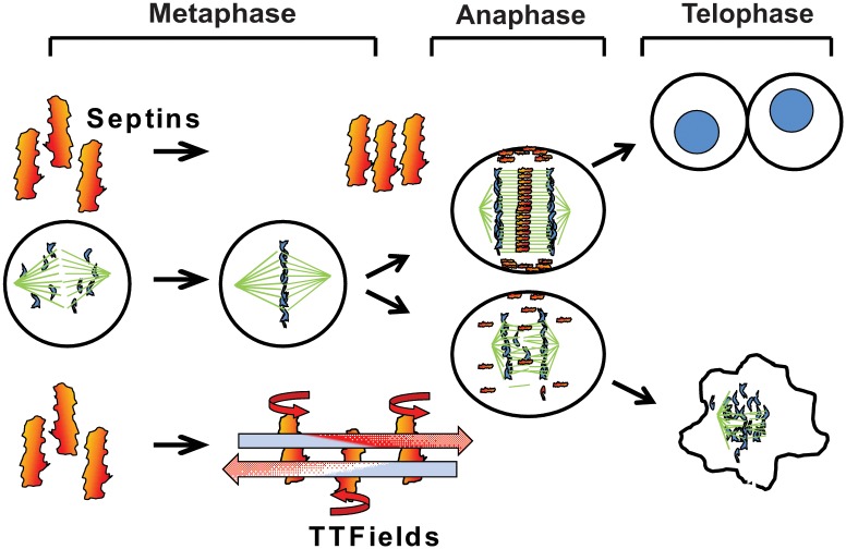
The anti-tumor effects of chemotherapy and radiation are thought to be mediated by triggering G1/S or G2/M cell cycle checkpoints, while spindle poisons, such as paclitaxel, block metaphase exit by initiating the spindle assembly checkpoint. In contrast, we have found that 150 kilohertz (kHz) alternating electric fields, also known as Tumor Treating Fields (TTFields), perturbed cells at the transition from metaphase to anaphase. Cells exposed to the TTFields during mitosis showed normal progression to this point, but exhibited uncontrolled membrane blebbing that coincided with metaphase exit. The ability of such alternating electric fields to affect cellular physiology is likely to be dependent on their interactions with proteins possessing high dipole moments. The mitotic Septin complex consisting of Septin 2, 6 and 7, possesses a high calculated dipole moment of 2711 Debyes (D) and plays a central role in positioning the cytokinetic cleavage furrow, and governing its contraction during ingression. We showed that during anaphase, TTFields inhibited Septin localization to the anaphase spindle midline and cytokinetic furrow, as well as its association with microtubules during cell attachment and spreading on fibronectin. After aberrant metaphase exit as a consequence of TTFields exposure, cells exhibited aberrant nuclear architecture and signs of cellular stress including an overall decrease in cellular proliferation, followed by apoptosis that was strongly influenced by the p53 mutational status. Thus, TTFields are able to diminish cell proliferation by specifically perturbing key proteins involved in cell division, leading to mitotic catastrophe and subsequent cell death.


Conflict of interest statement

Competing Interests: K. D. S. and E. T. W. received funding from NovoCure Inc., in the form of an unrestricted grant and travel expenses for training on the use of the equipment used in this study. E. T. W. was also one of the physicians who presented clinical trial data to an FDA panel in February 2011, prior to approval of the device. All research presented was conducted independently of the company, therefore these interactions do not alter the authors' adherence to PLOS ONE policies on sharing data and materials.

Figures

Fig 1
Fig 1. Cells exposed to TTFields exhibit an inability to progress through anaphase successfully resulting in cellular derangement.
MDA-MB-231 cells were synchronized using double aphidicolin block and then allowed to re-enter the cell cycle in the absence of TTFields (top panels), exposed to TTFields (middle panels) or treated with 3 μM paclitaxel (bottom panels). Cells were harvested and analyzed by FACS for transit through mitosis by staining with antibodies against Cyclin B antibodies and pH3 at 4, 7, 9, 11 and 13 hours after aphidicolin removal. Cells treated with TTFields did not exhibit a marked accumulation of mitotic cells showing a slight increased accumulation of cells at the 9 and 11 hour time points compared to control cultures suggesting a reduction in the rate of mitotic exit (A). Results are representative of 4 separate experiments. MDA-MB-231 cells were synchronized using aphidicolin followed by RO3306 treatment and were then allowed to progress through mitosis n the presence or absence of TTFields following drug removal. Cells were then collected at 60, 120 and 150 min and stained with antibodies against pH3 and 7-AAD for DNA content for analysis by FACS (B).
Fig 2
Fig 2. Cells exposed to TTFields during M-phase exhibit chromosomal disordering during the metaphase to anaphase progression.
HeLa cells were partially synchronized by treating with aphidicolin, then stained with DRAQ5 to visualize their chromosomes and subjected to fluorescence and phase contrast time lapse microscopy. Cells were imaged as they transited through mitosis either with or without TTFields exposure by both phase contrast and fluorescence during exposure to TTFields and time lapse series were captured. Single frames extracted from the time-lapse series of either Sham-treated (A) or TTFields-treated (B) cells visualized by phase contrast (left panels) or DRAQ5 (middle panels) at intervals of 240 seconds showed cells undergoing violent mitotic contractions that appeared coincident with the separation of daughter chromosomes at the onset of anaphase. Measurement of the time intervals between chromatid condensation and formation of the metaphase plate (C) and from the formation of the metaphase plate to either evidence of anaphase or TTFields-induced membrane contractions (D) were similar, 49.39 ± 1.988 min vs. 43.26 ± 2.088 min for plate formation and 47.04 ± 1.196 min vs. 44.36 ± 2.037 min for metaphase exit in Sham-treated vs TTFields-treated cultures, respectively. However, the outcome of mitosis was markedly different in Sham-treated (n = 130) vs. TTFields-treated (n = 85) cultures (E). Mitotic spindles and metaphase plates formed normally in TTFields treated cells (F), however, cells exiting mitosis in TTFields-treated cultures exhibited increased in abnormal nuclei with many cell possessing multiple micronuclear structures (G). Scale bar = 20 μm.
Fig 3
Fig 3. Septin 7 localization is perturbed by TTFields.
Crystal structure of Septin complex composed of Septins 2, 6, and 7 showing the direction of the dipole vector (2711Debyes) relative to the longitudinal axis of the heterotrimer (A). Synchronized cells were cultured in the absence of TTFields, TTFields delivered at 150 kHz (n = 30) or 500 kHz (n = 12), fixed and stained with antibodies against α-tubulin and Septin 7 and counter stained with DAPI. Confocal microscopy of cells captured in mid anaphase under these conditions revealed that Septin 7 localization was reduced and the midlines of anaphase spindles were perturbed by TTFields of 150 kHz but to a lesser extent at TTFields at 500 kHz compared to control cells (n = 27) (B). The average intensity of Septin 7 staining was separately determined within the region of the midline and for the entire cell. The ratio of the average intensity of Septin 7 staining at midline that of the total staining within the cell in Sham (n = 10) and presence of TTFields at 150kHz (n = 9) or 500kHz (n = 8) showed a significant decrease in Septin accumulation at the midlines in cells treated with 150 kHz TTFields (C). In contrast to Septin 7, PLK1 localization to the anaphase spindle midline appeared unperturbed by TTFields at 150 kHz (n = 11) (D). Scale bar = 10 μm. Septin organizes filamentous actin within the CCF during anaphase, in 150kHz TTFields treated cells F-actin accumulation were significantly decrease at the midline (E and F), n = 16 sham and n = 17 TTFields. To test whether TTFields interfered with the ability of Septins to re-localize to interphase microtubules during attachment and spreading on fibronectin, cells were allowed to spread for 9 hours under Sham conditions, or treated with TTFields at either 150 kHz, or 500 kHz (G). The Septin localization to the cytoskeleton was scored based on the striation of Septin 7 at the base of the cell with 1 equating to no diffuse staining with no striations, 2 equating to moderate localization and striations, and 3 equating to strong striations. While cells were allowed to re-attach and spread, cells in the absence of TTFields exposure exhibited strong Septin 7 association with microtubules (n = 70) while cells that were exposed to TTFields at 150 kHz (n = 152) exhibited significantly less Septin 7 localization to microtubules. Cell spreading while exposed to TTFields of 500 kHz (n = 104) exhibited an intermediate amount of microtubule association. Data was accumulated from three separate experiments (H). Scale bar = 50 μm.
Fig 4
Fig 4. Cells exposed to TTFields often result in mitotic disruption and subsequent cell death.
Upon their removal from the TTFields (right panel), cells exhibited significantly signs of cell stress including altered morphology compared to sham-treated controls (left panel) including increased size and vacularity (A). Insets show enlargement of cells in the field for detail. This increase in vacularity was also evident by increased side scatter by FACS (B). HeLa (upper panels) or MCF-7 cells (lower panels) were synchronized using aphidicolin, plated on gridded glass bottom dishes and either Sham-treated or exposed to TTFields during either mitosis or the G1 phase. After removal from the TTFields, cells were counted in individual grids at 4 and 24 hours after the termination of treatment and the resulting ratios were measured as a metric of proliferation. Most cells present at 4 hours remained at 24 hours. The proliferation of cells was significantly lower following exposure to TTFields during the M phase compared to sham-treated cultures. Both the TTFields-treated and Sham-treated cells exhibited similar proliferation when treated in G1 (C). HCT116 p53+/+ (upper panels) or HCT116 p53-/- cells (lower panels) were incubated for 24 hours either without (left panels) or with TTFields-exposure (right panels) and then incubated for an additional 24 hours. Cells were allowed to incorporate BrdU into their DNA as a measure of cells in S phase (D). To test if cells exposed to TTFields exhibited a higher incidence of apoptosis. HCT-116 p53+/+ cells were treated with TTFields for 24 hours and then further incubated at 37°C and then stained with FITC-labeled Annexin V at 18, 36, and 60 hours after the midpoint of their treatment. Annexin V binding to cells was visualized by fluorescence microscopy and scored for the presence of Annexin V positive cells. Cells were observed to undergo apoptosis after 18 hours of removal from TTFields with a peak at 36 hours (E). In order to test the effect of p53 depletion on TTFields-induced apoptosis, the responses of HCT-116 p53+/+ were compared with HCT116 p53-/- cells at 36 hours following TTFields treatment. p53+/+ with exposure to TTFields exhibited higher levels of apoptotic cells than their p53-/- counterparts (F).
Fig 5
Fig 5. Model for TTFields action leading to mitotic disruption.
During mitosis, the Septin 2, 6, 7 complex is recruited to the Anaphase spindle midline and the cytokinetic cleavage furrow by Anillin where it self-assembles into a fibrous lattice due to lateral interactions between parallel Septin filaments. By inducing rotational movement within the parallel fibers at a slightly less more than a right angle to their lateral axis, TTFields are able to inhibit the propagation of lattice formation by disrupting the ability of individual fibers to bind each other. In the absence of proper Septin function, contractile elements of the cytokinetic furrow are not restrained within the equatorial midline of the cell resulting in ectopic furrow malfunction that leads to violent membrane contractions at the onset of anaphase followed by aberrant mitotic exit.

References

    1. Sudakin V, Chan GK, Yen TJ. Checkpoint inhibition of the APC/C in HeLa cells is mediated by a complex of BUBR1, BUB3, CDC20, and MAD2. The Journal of cell biology. 2001;154(5):925–36. Epub 2001/09/06. 10.1083/jcb.200102093 - DOI - PMC - PubMed
    1. Kops GJ, Shah JV. Connecting up and clearing out: how kinetochore attachment silences the spindle assembly checkpoint. Chromosoma. 2012;121(5):509–25. Epub 2012/07/12. 10.1007/s00412-012-0378-5 . - DOI - PubMed
    1. Gruber S, Haering CH, Nasmyth K. Chromosomal cohesin forms a ring. Cell. 2003;112(6):765–77. Epub 2003/03/26. . - PubMed
    1. Haering CH, Farcas AM, Arumugam P, Metson J, Nasmyth K. The cohesin ring concatenates sister DNA molecules. Nature. 2008;454(7202):297–301. Epub 2008/07/04. 10.1038/nature07098 . - DOI - PubMed
    1. Oliveira RA, Hamilton RS, Pauli A, Davis I, Nasmyth K. Cohesin cleavage and Cdk inhibition trigger formation of daughter nuclei. Nature cell biology. 2010;12(2):185–92. Epub 2010/01/19. 10.1038/ncb2018 - DOI - PMC - PubMed

Publication types