Review
. 2015 May 22;6(2):267-98.
doi: 10.3390/genes6020267.

Replication stress in Mammalian cells and its consequences for mitosis

Affiliations
Review

Replication stress in Mammalian cells and its consequences for mitosis

Camille Gelot et al. Genes (Basel). .

Abstract

The faithful transmission of genetic information to daughter cells is central to maintaining genomic stability and relies on the accurate and complete duplication of genetic material during each cell cycle. However, the genome is routinely exposed to endogenous and exogenous stresses that can impede the progression of replication. Such replication stress can be an early cause of cancer or initiate senescence. Replication stress, which primarily occurs during S phase, results in consequences during mitosis, jeopardizing chromosome segregation and, in turn, genomic stability. The traces of replication stress can be detected in the daughter cells during G1 phase. Alterations in mitosis occur in two types: 1) local alterations that correspond to breaks, rearrangements, intertwined DNA molecules or non-separated sister chromatids that are confined to the region of the replication dysfunction; 2) genome-wide chromosome segregation resulting from centrosome amplification (although centrosomes do not contain DNA), which amplifies the local replication stress to the entire genome. Here, we discuss the endogenous causes of replication perturbations, the mechanisms of replication fork restart and the consequences for mitosis, chromosome segregation and genomic stability.

Keywords: anaphase bridges; centrosome; fragile sites; homologous recombination; micronuclei; mitosis; replication stress; single-ended DSB.


Figures

Figure 1
Figure 1
Double-strand break repair models that act via homologous recombination (HR). Left panel: Gene conversion. After resection, the single-stranded 3' tail invades a homologous, intact double-stranded DNA, forming a d-loop (displacement loop). This process tolerates a limited number of imperfect sequence homologies, thus creating heteroduplex intermediates bearing mismatches (yellow circles). The invading 3'-end primes DNA synthesis, which then fills in the gaps. The cruciform junctions (Holliday junctions, HJ) migrate. Resolution (or dissolution) of HJs occurs in two different orientations (orange or red triangles), resulting in gene conversion either with or without crossing over. Middle panel: Break-induced replication (BIR). The initiation is similar to that of the previous models, but the synthesis continues over longer distances on the chromosome arms, even reaching the end of the chromosome. Here, there is neither resolution of the HR nor crossover. Right panel: Synthesis-dependent strand annealing (SDSA). Initiation is similar to that of the previous model, but the invading strand de-hybridizes and re-anneals at the other end of the injured molecule; no HJ is formed.
Figure 2
Figure 2
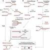
Fork restarts by HR following replication stress. (A) Model of repair of blocking lesions. (A.1) DNA adducts obstruct DNA synthesis by replicative DNA polymerases. Fork progression on a damaged template might involve a repriming event downstream of the damage, which leaves a ssDNA gap behind the moving fork. Rad51 then nucleates on the ssDNA gaps and promotes the recombination with the sister chromatid to seal the gap. Other mechanisms might be involved in the bypass of DNA lesions such as translesion synthesis (TLS). (A.2) Model of fork regression at a stalled fork: A slowing down of fork velocity or fork arrest leads to a transient uncoupling of the helicase and polymerases, thus exposing ssDNA at the stalled fork. The fork reversion forms a “chicken foot” structure (i.e., the fork and the nascent strand, which is complementary, being annealed together to form a four-way junction). Cleavage of this structure might involve MUS81 and leads to single-ended DSB formation. (B) Model of broken-fork repair. A replication fork can be converted into single-ended DSBs following the passage of the fork through a nick or following cleavage by an endonuclease. The single-ended break is then resected and Rad51 nucleates on the exposed ssDNA and promotes recombination with the sister chromatid. The 3' end of the invading strand primes DNA synthesis, and the replisome has been proposed to be rebuilt from the extended d-loop structure. (C) Model of fork restarts at a collapsed fork. Fork collapse might arise from a stalled fork where the replisome fails to be maintained in a functional state or when the replisome encounters physical obstacles such as tightly DNA bound proteins or RNA/DNA hybrids. Resection of nascent strands might help the fork to regress (i.e., the fork moving backward without the annealing of nascent strands) and thus allow the 3' end of the nascent strand to be extruded. Rad51 nucleates on the exposed ssDNA and promotes recombination with the parental DNA duplex. The replisome could again be rebuilt from the extended d-loop.
Figure 3
Figure 3
Joining of single-ended double strand breaks (DSBs) could lead to rearrangements. (Left panel): A single-ended DSB generated by replication stress is normally repaired by SCE in a conservative way. Rearrangements occur when a single-ended DSB is joined to another single-ended DSB, which is likely to be distal. (Middle panel): The annealing of few nucleotides at the extremity of the single ended DSB with another broken fork activates the MMBIR (microhomology-mediated break-induced replication) mechanism. MMBIR coupled to several switches in fork templates leads to complex rearrangements and has been proposed to be a mechanism that originates chromotripsis. (Right panel): The end-joining (EJ) by C-NHEJ or A-EJ of the single ended DSB with another single-ended DSB lead to dicentric chromosome formation.
Figure 4
Figure 4
DSB repair pathway models. (Left panel): Canonical C-NHEJ. The heterodimer Ku80-Ku70 binds to DNA ends, which then recruits DNA-PKcs. In subsequent steps, several proteins including Artemis, polynucleotide kinase (PNK), and members of the polymerase X family process the DNA ends. In the last step, ligase IV associated with its co-factors Xrcc4 and Cernunos/XLF joins the ends (for review about cNHEJ and A-EJ actors see [148,151]). (Right Panel): Resection as a common initiation step for HR and A-EJ at DSB. 53BP1, RIF1 and Ku70-80 heterodimer protect DSB ends from resection and HR and A-EJ actions. The CDK1/2-dependent phosphorylation of CtIP and EXO1 favors the initiation of resection and extension, respectively [95,103,152]. Recently, REV7/MAD2L2 was described as an inhibitor of resection and HR, although its role in A-EJ inhibition was not directly studied and remains hypothetical [104,105]. A short ssDNA resection allows for A-EJ but not homologous recombination, while a long ssDNA resection allows for both A-EJ and HR; however, HR requires the presence of homologous sequences. Recently, POLQ polymerase was shown to inhibit HR and to promote A-EJ at DSBs [153,154]. A-EJ results in repair that is error-prone and is associated with deletions at the repair junctions with frequent use of microhomologies that are distant from the DSB. Alternative-EJ: Parp1 plays a role in the initiation process, and it has been proposed that a single-strand DNA resection reveals complementary microhomologies (two to four nucleotides or more in length) that can anneal, with gap-filling completing the end-joining. A-EJ is always associated with deletions at the junctions and can involve microhomologies (MMEJ or microhomologies-mediated EJ) that are distant from the DSB. Subsequently, Xrcc1 and ligase III (which can be substituted by ligase I) complete the A-EJ process. Homologous recombination: The first step, which is the initiation of resection, involves the removal of ~50–100 bases of DNA from the 5' end by the MRN complex (Mre11-Rad50-Nbs1) in conjunction with CtIP. The second step, resection extension, is carried out by two alternate pathways involving either the 5' to 3' exonuclease EXO1 or the helicase-topoisomerase complex BLM-TOPIIIα-RMI1-2 in concert with the nuclease CtIP/DNA2. WRN helicase has also been shown to act with CtIP and to stimulate resection in human cells [155].
Figure 5
Figure 5
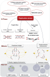
Replicative stress and its consequences in mitosis. Replication stress from either endogenous or exogenous causes (red circles) leaves chromosomal segment unreplicated or interwinded leading to anaphase bridges formation. Single-ended DSB could lead to dicentric chromosome formation and thus, also, to anaphase bridge formation. Non-detected damages upon low replicative stress could be grouped in one detectable entity in G1: 53BP1 bodies and/or micronuclei. Replication stress also favors mitotic extra centrosomes and multipolar mitosis, thus amplifying mitotic catastrophes and genome instability to the whole genome.

References

    1. Gorgoulis V.G., Vassiliou L.F., Karakaidos P., Zacharatos P., Kotsinas A., Liloglou T., Venere M., Ditullio R.A., Jr., Kastrinakis N.G., Levy B., et al. Activation of the DNA damage checkpoint and genomic instability in human precancerous lesions. Nature. 2005;434:907–913. doi: 10.1038/nature03485. - DOI - PubMed
    1. Bartkova J., Horejsí Z., Koed K., Krämer A., Tort F., Zieger K., Guldberg P., Sehested M., Nesland J.M., Lukas C., et al. DNA damage response as a candidate anti-cancer barrier in early human tumorigenesis. Nature. 2005;434:864–870. doi: 10.1038/nature03482. - DOI - PubMed
    1. Burrell R.A., McClelland S.E., Endesfelder D., Groth P., Weller M.-C., Shaikh N., Domingo E., Kanu N., Dewhurst S.M., Gronroos E., et al. Replication stress links structural and numerical cancer chromosomal instability. Nature. 2013;494:492–496. doi: 10.1038/nature11935. - DOI - PMC - PubMed
    1. Flach J., Bakker S.T., Mohrin M., Conroy P.C., Pietras E.M., Reynaud D., Alvarez S., Diolaiti M.E., Ugarte F., Forsberg E.C., et al. Replication stress is a potent driver of functional decline in ageing haematopoietic stem cells. Nature. 2014;512:198–202. doi: 10.1038/nature13619. - DOI - PMC - PubMed
    1. Anglana M., Apiou F., Bensimon A., Debatisse M. Dynamics of DNA replication in mammalian somatic cells: Nucleotide pool modulates origin choice and interorigin spacing. Cell. 2003;114:385–394. doi: 10.1016/S0092-8674(03)00569-5. - DOI - PubMed