. 2015 May 27:8:33.
doi: 10.1186/s13041-015-0122-1.

Full UPF3B function is critical for neuronal differentiation of neural stem cells

Affiliations

Full UPF3B function is critical for neuronal differentiation of neural stem cells

Tahani Alrahbeni et al. Mol Brain. .

Abstract

Background: Mutation in the UPF3B gene on chromosome X is implicated in neurodevelopmental disorders including X-linked intellectual disability, autism and schizophrenia. The protein UPF3B is involved in the nonsense-mediated mRNA decay pathway (NMD) that controls mRNA stability and functions in the prevention of the synthesis of truncated proteins.

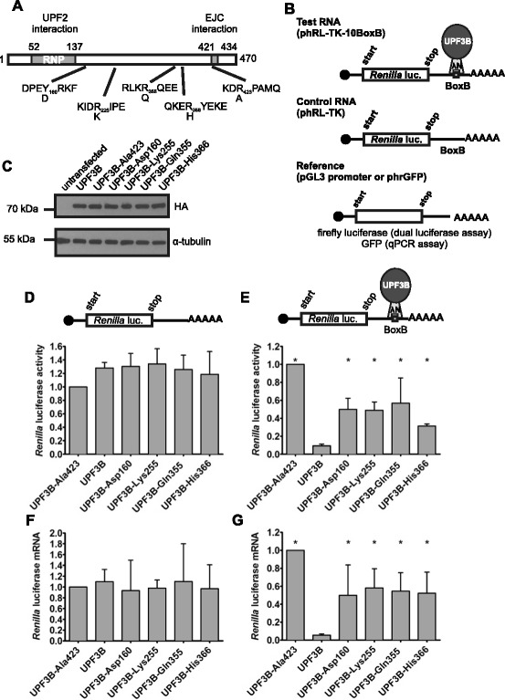
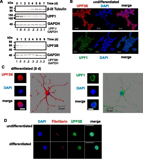
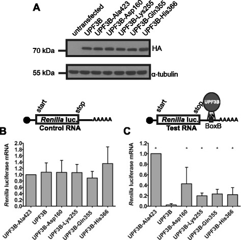
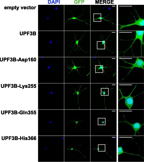
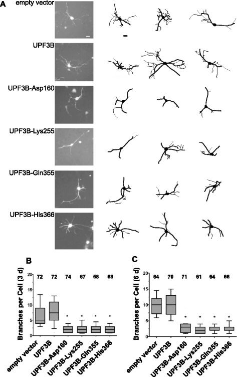
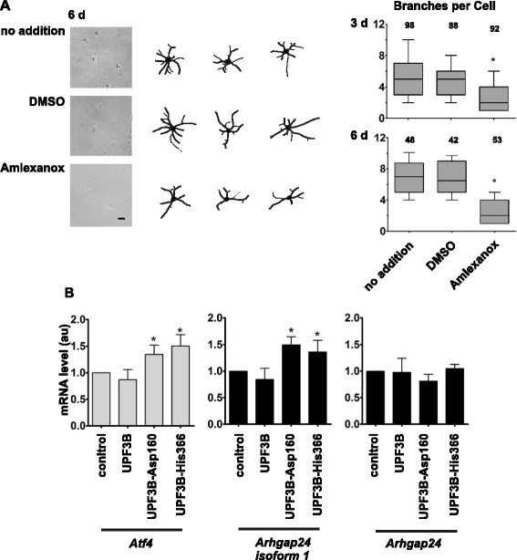
Results: Here we show that NMD pathway components UPF3B and UPF1 are down-regulated during differentiation of neural stem cells into neurons. Using tethered function assays we found that UPF3B missense mutations described in families with neurodevelopmental disorders reduced the activity of UPF3B protein in NMD. In neural stem cells, UPF3B protein was detected in the cytoplasm and in the nucleus. Similarly in neurons, UPF3B protein was detected in neurites, the somatic cytoplasm and in the nucleus. In both cell types nuclear UPF3B protein was enriched in the nucleolus. Using GFP tagged UPF3B proteins we found that the missense mutations did not affect the cellular localisation. Expression of missense mutant UPF3B disturbed neuronal differentiation and reduced the complexity of the branching of neurites. Neuronal differentiation was similarly affected in the presence of the NMD inhibitor Amlexanox. The expression of mutant UPF3B proteins lead to a subtle increase in mRNA levels of selected NMD targets.

Conclusions: Together our findings indicate that, despite the down-regulation of NMD factors, functional NMD is critical for neuronal differentiation. We propose that the neurodevelopmental phenotype of UPF3B missense mutation is caused by impairment of NMD function altering neuronal differentiation.


Figures

Fig. 1
Fig. 1
Missense mutations impair UPF3B activity in NMD. a. Schematic representation of UPF3B protein structure with location of protein-protein interaction domains and amino acid changes. N-terminal N peptide and HA tag sequences are not shown. b. Schematic representation of the tethering assay mimicking NMD. λNUPF3B protein is tethered to Renilla luciferase mRNA with BoxB elements in the 3’ UTR expressed from phRL-TK-10BoxB (Test RNA) but is unable to bind mRNA expressed from phRL-TK lacking BoxB elements (Control RNA). Co-transfected pGL3-promoter or phrGFP serve as standards in dual luciferase and qPCR assays, respectively (Reference). c. Expression of λN-HA-UPF3B proteins. HeLa cells were transfected with pCI-λN-HA-UPF3B expression constructs and lysed after 48 h. Expression of UPF3B proteins and α-tubulin was analysed by 10 % SDS PAGE followed by Western blotting with anti-HA and anti-tubulin antibodies. d, e. Tethering assay: Luciferase activity. Hela cells were transfected with phRL-TK (d) or phRL-TK-10BoxB (e), together with reference plasmid pGL3-promoter and the pCI-λN-HA-UPF3B expression constructs. Luciferase activities were measured 48 h after transfection. Renilla luciferase activities were standardised with respect to firefly luciferase activity, and the activity in cells expressing UPF3B-Ala423 was defined as 1. Shown are average Renilla luciferase activities from three independent experiments. f, g: Tethering assay: Luciferase mRNA levels. HeLa cells were co-transfected with phRL-TK (f) or phRL-TK-10BoxB (g) but with phrGFP instead of pGL3-promoter. RNA was prepared 48 h later and mRNA levels were determined by qPCR. Luciferase mRNA levels were standardised with respect to GFP mRNA levels, and luciferase mRNA in cells expressing UPF3B-Ala423 was defined as 1. Shown are average luciferase mRNA levels from four independent experiments. Error bars indicate standard deviations, asterisks indicate values significantly different from luciferase activity or mRNA levels in the presence of UPF3B (one-way ANOVA followed by Dunnett's test; P < 0.05).
Fig. 2
Fig. 2
NMD factors UPF1 and UPF3 localise to neurites while protein levels are downregulated during neuronal differentiation. a. Neural stem cells were differentiated by the addition of forskolin and retinoic acid. Protein samples were prepared before (day 0 (d 0)) and at the indicated time (d, days) after induction of differentiation. Equal amounts of protein were subjected to 8 % SDS PAGE and analysed for β-III Tubulin, UPF3B and UPF1 protein levels by Western blotting. Samples were probed for GAPDH protein as loading control. Proteins with faster mobility indicated by asterisk reflect the behaviour of UPF1 and UPF3B and are likely to be alternate isoforms. The relative changes of UPF3B and UPF1 protein levels were quantitated using GAPDH as reference, with levels at day 0 defined as 1. The mobility of relevant molecular mass markers is indicated. b. UPF1 and UPF3B protein localisation in undifferentiated HCN-A94 cells. Cells were stained with DAPI (blue) and with either anti-UPF3B antibody (red) or anti-UPF1 antibody (green) were analysed by confocal microscopy. Shown are separate channels and merged images from a single section. c. UPF3B and UPF1 localisation in neurons. Neural stem cells differentiated for 8 days and then stained with DAPI and either anti-UPF3B antibody or anti-UPF1 antibody were analysed by confocal microscopy. Shown are separate channels and merged images from a single section through the nucleus and 3 days renderings of corresponding Z-stacks. The scale bars represent 20 μm. Note that UPF3B and UPF1 are detected in neurites and UPF1, but not UPF3B, largely is excluded from the nucleus. d. Nucleolar localisation of UPF3B. Undifferentiated or for 8 day differentiated neural stem cells were stained with anti-UPF3B antibodies, anti-Fibrillarin antibodies and DAPI and then analysed by confocal microscopy. Sections shown focus on the nuclear compartment. The scale bars represent 20 μm. Note that UPF3B is enriched in nucleoli stained by anti-Fibrillarin antibodies
Fig. 3
Fig. 3
Mutations in UPF3B impair UPF3B protein NMD activity in neural stem cells. a. Expression of λN-HA-UPF3B proteins. Neural stem cells were transfected with pCI-λN-HA-UPF3B expression constructs and protein expression was analysed 48 h after transfection by 10 % SDS PAGE followed by detection by Western blotting. UPF3B proteins and α-tubulin were detected using anti-HA and anti-tubulin antibodies. b, c. Tethering assays. b. Neural stem cells were transfected with the control plasmid phRL-TK, the reference plasmid phrGFP-C and the various pCI-λN-HA-UPF3B expression constructs. 24 h after transfection, differentiation was induced. RNA was prepared 48 h after transfection and analysed by qPCR as described. c. Neural stem cells were transfected, treated and mRNA levels analysed as in B except that phRL-TK was replaced with phRL-TK-10BoxB. For the graphs, Renilla luciferase mRNA levels were standardised with respect to GFP mRNA levels, and Renilla luciferase mRNA levels in cells expressing UPF3B-Ala423 was defined as 1. The graphs show average Renilla luciferase mRNA levels from three independent experiments, error bars indicate the standard deviation. Asterisks indicate values significantly different from luciferase mRNA levels in the presence of UPF3B (one-way ANOVA followed by Dunnett's test; P < 0.05)
Fig. 4
Fig. 4
Mutations in UPF3B do not affect its cellular distribution in neurons. Neural stem cells were transfected with the pEGFP-C3 expressing GFP only (empty vector) or a pEGFP-C3 derivative expressing one of the UPF3B forms with an N-terminal GFP tag. Cells were diluted and re-plated after 24 h, and then differentiated for 6 days prior to analysis by confocal microscopy. Shown are separate Z-stack projections of GFP and DAPI channels and of the merged channels. The origin region of the enlarged section is indicated. The scale bar represents 20 μm
Fig. 5
Fig. 5
Expression of mutant UPF3B proteins affects neuronal differentiation of neural stem cells. Neural stem cells were transfected with pIRES-GFP (empty vector) or pIRES-GFP derivatives expressing the indicated UPF3B proteins. Differentiation was induced after 24 h, and analysed after 3 days and 6 days of differentiation by microscopy. Transfected cells were identified and visualised by GFP fluorescence. The analysis of neuron differentiation was performed as described in Methods. a. Representative pictures of neurons with tracing, and tracing of 2 additional neurons after 6 days of differentiation illustrating a reduction in branching in neurons expressing mutant UPF3B proteins. The scale bars represent 20 μm. Tracings are to scale with primary neurites traced with thicker lines then branches protruding from primary neurites. b, c. Branching was analysed after 3 days and 6 days as described and results are presented as box plots. The box plots show the median, with boxes spanning 25th- 75th percentiles and the crossbars indicating the 10th and 90th percentiles. Experiments were done independently 3 times with similar outcomes and the data were combined for presentation. Numbers of cells analysed are indicated. Asterisks indicate significantly reduced branching in neurons expressing mutant UPF3B proteins compared to neurons transfected with the empty vector (Kruskal Wallis test followed by Dunn's multiple comparison test; P < 0.05)
Fig. 6
Fig. 6
A functional NMD pathway is important for neuronal differentiation. a. Treatment with NMD inhibitor Amlexanox affects neuronal differentiation of neural stem cells. Neural stem cells were differentiated in the presence of DMSO or 10 μM Amlexanox or without any further addition. Neurons were analysed 3 days and 6 days after induction of differentiation as described. Shown are representative pictures of neurons with tracing, and tracing of 2 additional neurons after 6 days of differentiation illustrating a reduction in branching in Amlexanox treated neurons. The scale bar represents 20 μm. Results are plotted showing the median, with boxes spanning 25th- 75th percentiles and the crossbars indicating the 10th and 90th percentiles. Experiments were done independently 3 times with similar outcomes and the data were combined for presentation. Numbers of cells analysed are indicated. The Asterisk indicates that branching of Amlexanox treated neurons is significantly reduced compared to neurons differentiated without addition any addition or DMSO only (Kruskal Wallis test followed by Dunn's multiple comparison test; P < 0.05). b. Expression of mutant UPF3B proteins that affect neuronal differentiation causes an increase in mRNA levels of selected NMD substrates. Neural stem cells were transduced with lentivirus without insert (control) or expressing either UPF3B, UPF3B-Asp160 or UPF3B-His366 protein. 24 h after transduction, differentiation was induced. RNA was isolated after six days of differentiation and gene expression was analysed by reverse transcription followed by quantitative PCR. mRNA levels of NMD substrates Atf4 and Arhgap24 isoform 1, and of transcripts encoding all shorter Arhgap24 isoforms not subject to NMD control were measured and standardised using Gapdh mRNA as reference. Atf4 and Arhgap24 mRNA levels in cells transduced with the control virus were set as 1. Asterisks indicate values significantly increased compared to levels in cells transduced with the control virus (one-way ANOVA followed by Dunnett's test; P < 0.05)

References

    1. Addington AM, Gauthier J, Piton A, Hamdan FF, Raymond A, Gogtay N, et al. A novel frameshift mutation in UPF3B identified in brothers affected with childhood onset schizophrenia and autism spectrum disorders. Mol Psychiatry. 2010;16:238–239. doi: 10.1038/mp.2010.59. - DOI - PMC - PubMed
    1. Tarpey PS, Lucy Raymond F, Nguyen LS, Rodriguez J, Hackett A, Vandeleur L, et al. Mutations in UPF3B, a member of the nonsense-mediated mRNA decay complex, cause syndromic and nonsyndromic mental retardation. Nat Genet. 2007;39:1127–1133. doi: 10.1038/ng2100. - DOI - PMC - PubMed
    1. Laumonnier F, Shoubridge C, Antar C, Nguyen LS, Van Esch H, Kleefstra T, et al. Mutations of the UPF3B gene, which encodes a protein widely expressed in neurons, are associated with nonspecific mental retardation with or without autism. Mol Psychiatry. 2010;15:767–776. doi: 10.1038/mp.2009.14. - DOI - PubMed
    1. Lynch SA, Nguyen LS, Ng LY, Waldron M, McDonald D, Gecz J. Broadening the phenotype associated with mutations in UPF3B: Two further cases with renal dysplasia and variable developmental delay. Eur J Med Genet. 2012;55:476–479. doi: 10.1016/j.ejmg.2012.03.010. - DOI - PubMed
    1. Szyszka P, Sharp SI, Dedman A, Gurling HM, McQuillin A. A nonconservative amino acid change in the UPF3B gene in a patient with schizophrenia. Psychiatr Genet. 2012;22:150–151. doi: 10.1097/YPG.0b013e32834accbe. - DOI - PubMed

Publication types