. 2016 Jun;36(6):837-46.
doi: 10.1111/liv.12876. Epub 2015 Jun 22.

Bile duct-ligated mice exhibit multiple phenotypic similarities to acute decompensation patients despite histological differences

Affiliations

Bile duct-ligated mice exhibit multiple phenotypic similarities to acute decompensation patients despite histological differences

Alastair O'Brien et al. Liver Int. 2016 Jun.

Abstract

Background & aims: Patients with decompensated cirrhosis are susceptible to infection. Innate immune dysfunction and development of organ failure are considered to underlie this. A rodent model of liver disease sharing these phenotypic features would assist in vivo study of underlying mechanisms and testing of therapeutics. We evaluated three models to identify which demonstrated the greatest clinical and immunological phenotypic similarity to patients with acutely decompensated (AD) cirrhosis.

Methods: We selected Bile Duct Ligation (BDL) rats at 4 weeks, BDL mice at 14 days and Carbon tetrachloride (CCl4 ) mice at 10 weeks (with studies performed 7 days after final CCl4 infection). We examined organ dysfunction, inflammatory response to carrageenan-in-paw, plasma eicosanoid concentrations, macrophage cytokine production and responses to peritoneal infection.

Results: Bile duct ligation caused sarcopenia, liver, cardiovascular and renal dysfunction whereas CCl4 mice demonstrated no clinical abnormalities. BDL rodents exhibited depressed response to carrageenan-in-paw unlike CCl4 mice. BDL rats have slightly elevated plasma eicosanoid levels and plasma showed partial PGE2 -mediated immune suppression whereas CCl4 mice did not. Plasma NOx was elevated in patients with acute or chronic liver failure (AoCLF) compared to healthy volunteers and BDL rodents but not CCl4 mice. Elevated nitric oxide (NO) via inducible nitric oxide synthase (iNOS) mediates defective leucocyte trafficking in BDL rodent models.

Conclusions: We conclude that BDL mice and rats are not simply models of cholestatic liver injury but may be used to study mechanisms underlying poor outcome from infection in AD and have identified elevated NO as a potential mediator of depressed leucocyte trafficking.

Keywords: bile duct ligation; carbon tetrachloride; eicosanoids; immune suppression; leucocyte trafficking.


Figures

Figure 1
Figure 1
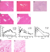
(a‐i) Haematoxylin and Eosin (HE)‐stained histological sections of the portal tract of liver from bile duct ligated (BDL) rats showing ductular reaction and scant inflammation. (a‐ii) HE‐stained histological sections from Sham rats (28 days post procedure). (a‐iii) Elastica Van Gieson (EVG) staining of BDL rat liver demonstrating bridging fibrosis. (b‐i) HE‐stained histological sections of the portal tract of liver from CCl4 mice (at 10 weeks, samples taken one week after final CCl4 injection) showing nodular parenchyma with no inflammation and (b‐ii) sham mice. (c) Effect of carrageenan‐induced paw oedema (1%, 50 μl intra plantar injection in mice, 100 μl in rats) in (i) BDL mice (14 days post procedure, n = 9), (ii) CCl4 mice (n = 6) and (iii) BDL in rats (n = 6). Graphs show difference in paw size between carrageenan‐injected and saline‐injected paw. (d) Representative sections through rat paw 4 h following carrageenan from (i) sham and (ii) BDL animals. All histology slides at magnification ×20. For carrageenan experiments 3–6 animals per group were used. Data are represented as mean ± SEM. ** < 0.01 ***< 0.001, anova.
Figure 2
Figure 2
(a) Lipidomic analysis demonstrated that (i) PGE 2 and (ii) D2 were minimally elevated in BDL rats compared to shams. (b) Plasma from BDL rats induced a partial reduction in (i) LPS‐induced TNFα production from naïve mice peritoneal macrophages compared to plasma from shams which was attenuated when the EP1–3/DP selective inhibitor AH6809 was added in vitro or if plasma from BDL rats treated with indomethacin (Indo) was added. (b) Plasma from BDL rats had no effect on (ii) IL10 secretion from naïve mice peritoneal macrophages. (c) (i) PGE 2 and (ii) iD 2 levels were minimally elevated in plasma from Chronic CCl4 mice but (d) this plasma had no effect on peritoneal macrophage LPS‐induced (i) TNFα or (ii) IL10 production function compared to sham plasma.
Figure 3
Figure 3
(a‐i) Plasma nitrite levels in patients admitted to hospital with complications of cirrhosis (CP) compared to healthy volunteers (HV). (a‐ii) Plasma NOx levels in sham and liver injury rodents (n = 6 for each group). (b) Total leucocyte count following zymosan‐induced peritonitis in naïve, sham & BDL mice ± zymosan (Z; 0.1 mg at 4 h) and BDL mice warmed (warm) to the same core temperature as shams (b‐i) as well as CCl4 mice (b‐ii); n = 8–10 – data taken from 3 consecutive experiments. (c) Representative flow cytometry traces of GR1 and F4/80 labelled peritoneal macrophages 4 h after zymosan in naïve, sham, BDL and BDL + LNAME mice. These cells are gated on CD3 negative CD19 negative and CD 11B positive to exclude T and B cells. (d‐i and ii) Zymosan (0.1 mg) was injected, i.p. to naïve, sham and BDL mice with/without NOS inhibition [1400W (iNOS inhibitor), L‐NAME], with peritoneal PMNs showing the greatest reduction in cell numbers with full reversal seen in the presence of NOS inhibition. (d‐iii) Colony forming units detected in sham or BDL mouse blood at 3 h following i.p. injection of Group B streptococcus (GBS, 30 million units per mouse). BDL mice were treated with or L‐NAME (50 mg/kg) 30 mins before bacteria injection. Sham and BDL data as previously shown10, reproduced for this figure as experiments performed using L‐NAME were performed together to minimize animal usage. *< 0.05 **< 0.01 ***< 0.001, anova or t‐test where appropriate.

References

    1. Blachier M, Leleu H, Peck‐Radosavljevic M, Valla DC, Roudot‐Thoraval F. The burden of liver disease in Europe: a review of available epidemiological data. J Hepatol 2013; 58: 593–608. - PubMed
    1. Zatonski WA, Sulkowska U, Manczuk M, et al Liver cirrhosis mortality in Europe, with special attention to Central and Eastern Europe. Eur Addict Res 2010; 16: 193–201. - PubMed
    1. Navasa M, Fernandez J, Rodes J. Bacterial infections in liver cirrhosis. Ital J Gastroenterol Hepatol 1999; 31: 616–25. - PubMed
    1. Hassner A, Kletter Y, Shlag D, et al Impaired monocyte function in liver cirrhosis. Br Med J (Clin Res Ed) 1981; 282: 1262–3. - PMC - PubMed
    1. Fierer J, Finley F. Deficient serum bactericidal activity against Escherichia coli in patients with cirrhosis of the liver. J Clin Invest 1979; 63: 912–21. - PMC - PubMed

Publication types

LinkOut - more resources