. 2015 May 27:13:49.
doi: 10.1186/s12958-015-0047-7.

Uterine NDRG2 expression is increased at implantation sites during early pregnancy in mice, and its down-regulation inhibits decidualization of mouse endometrial stromal cells

Affiliations

Uterine NDRG2 expression is increased at implantation sites during early pregnancy in mice, and its down-regulation inhibits decidualization of mouse endometrial stromal cells

Yan Gu et al. Reprod Biol Endocrinol. .

Erratum in

Abstract

Background: N-myc down-regulated gene 2 (NDRG2) is a tumor suppressor involved in cell proliferation and differentiation. The aim of this study was to determine the uterine expression pattern of this gene during early pregnancy in mice.

Methods: Uterine NDRG2 mRNA and protein expression levels were determined by RT-PCR and Western blot analyses, respectively, during the peri-implantation period in mice. Immunohistochemical (IHC) analysis was performed to examine the spatial localization of NDRG2 expression in mouse uterine tissues. The in vitro decidualization model of mouse endometrial stromal cells (ESCs) was used to evaluate decidualization of ESCs following NDRG2 knock down by small interfering RNA (siRNA). Statistical significance was analyzed by one-way ANOVA using SPSS 19.0 software.

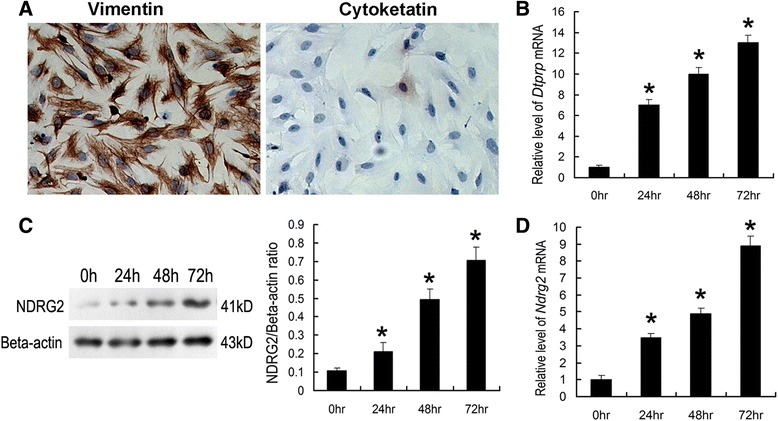
Results: Uterine NDRG2 gene expression was significantly up-regulated and was predominantly localized to the secondary decidual zone on days 5 and 8 of pregnancy in mice. Its increased expression was associated with artificial decidualization as well as the activation of delayed implantation. Furthermore, uterine NDRG2 expression was induced by estrogen and progesterone treatments. The in vitro decidualization of mouse ESCs was accompanied by up-regulation of NDRG2 expression, and knock down of its expression in these cells by siRNA inhibited the decidualization process.

Conclusions: These results suggest that NDRG2 might play an important role in the process of decidualization during early pregnancy.


Figures

Fig. 1
Fig. 1
NDRG2 expression in the mouse uterus during the estrous cycle. Upper: immunohistochemical analysis of NDRG2 expression during the diestrus (a), proestrus (b), estrous (c) and metestrus (d) phases in the mouse uterus. Below: quantitative PCR analysis of NDRG2 mRNA expression (e) in the uterus during the estrous cycle (n = 3). The thick arrow shows the luminal epithelium. The small arrow indicates the glandular epithelium. The columns with different superscripts are significantly different (P < 0.05)
Fig. 2
Fig. 2
NDRG2 expression in the mouse uterus during early pregnancy. a Quantitative PCR analysis of NDRG2 mRNA expression in the uterus during early pregnancy (n = 3, *P < 0.05). b Western blot and densitometric analyses of uterine NDRG2 protein levels during early pregnancy (n = 3). All experiments were repeated three times. The data are shown as the mean ± SEM. *P < 0.05. c Immunohistochemical analysis of uterine NDRG2 protein expression on days 1, 3, 4, 5, and 8 of pregnancy. IS, implantation sites; NI, non-implantation sites. *, indicates the location of the embryo. le, luminal epithelium; ge, glandular epithelium; st, stroma; pdz, primary decidual zone; sdz, secondary decidual zone; D, day of pregnancy; gc, giant cell; Scale bar represents 100 μm
Fig. 3
Fig. 3
The effects of steroid hormones on NDRG2 expression in the uterus. Upper: immunohistochemical analysis of NDRG2 protein expression in the uteri of ovariectomized mice after oil (a), progesterone (b), estrogen (c), and progesterone plus estrogen treatments (d). Below: quantitative PCR analysis of NDRG2 mRNA expression (e) in the uterus following steroid hormone treatment (n = 3). The thick arrow indicates the luminal epithelium. The small arrow shows the glandular epithelium. The columns with different superscripts are significantly different (P < 0.05)
Fig. 4
Fig. 4
Uterine NDRG2 expression following artificial decidualization and activation of delayed implantation. Immunohistochemical analysis of uterine NDRG2 protein expression following artificial decidualization (b) and in its contralateral uninjected uterine horn (a), under delayed implantation (d) and activation (e). Quantitative PCR analysis of NDRG2 mRNA expression in the uterus following artificial decidualization (c) and activation of delayed implantation (f) (n = 3). The thick arrow indicates the luminal epithelium. The small arrow shows the glandular epithelium. De, decidua; *, significantly different from control (P < 0.05)
Fig. 5
Fig. 5
In vitro decidualization of mouse primary ESCs. ESCs isolated from the uteri of day 4 pregnant mice were cultured in the presence of P4 and E2. a Immunocytochemical detection of vimentin and cytokeratin in cultured ESCs. b Quantitative PCR analysis of DTPRP mRNA expression in ESCs cultured for up to 72 h. c Western blot analysis of NDRG2 protein expression in ESCs cultured for up to 72 h. Densitometric analyses of NDRG2 at each time point compared with 0 h is shown. d Quantitative PCR analysis of NDRG2 mRNA expression in cultured ESCs. The relative fold induction of NDRG2 mRNA expression at each time point compared with its expression in the 0 h sample is shown. The values represent the mean ± SEM, as determined from three separate experiments. *, significantly different (P < 0.05)
Fig. 6
Fig. 6
Down-regulation of NDRG2 expression in ESCs inhibits in vitro decidualization. Cultured mouse ESCs were transfected with NDRG2-targeting siRNAs (100 nM) (a non-targeting siRNA was used as a control) at the time of plating. Quantitative PCR analyses of NDRG2 mRNA expression (a) and DTPRP mRNA expression (b) in ESCs transfected with NDRG2-targeting siRNAs or non-targeting siRNAs at 24 h and 48 h. Western blot (c) and densitometric analyses (d) of NDRG2 protein levels in ESCs at 72 h after transfection. The relative fold induction of NDRG2 protein expression compared with its expression in non-siRNA-treated group is shown. The values represent the mean ± SEM, as determined from three separate experiments. *, significantly different (P < 0.05)

Similar articles

Cited by

References

    1. Aplin JD. Embryo implantation: the molecular mechanism remains elusive. Reprod Biomed Online. 2006;13(6):833–9. doi: 10.1016/S1472-6483(10)61032-2. - DOI - PubMed
    1. Harun R, Ruban L, Matin M, Draper J, Jenkins NM, Liew GC, et al. Cytotrophoblast stem cell lines derived from human embryonic stem cells and their capacity to mimic invasive implantation events. Hum Reprod. 2006;21(6):1349–58. doi: 10.1093/humrep/del017. - DOI - PubMed
    1. Ferretti C, Bruni L, Dangles-Marie V, Pecking AP, Bellet D. Molecular circuits shared by placental and cancer cells, and their implications in the proliferative, invasive and migratory capacities of trophoblasts. Hum Reprod Update. 2007;13(2):121–41. doi: 10.1093/humupd/dml048. - DOI - PubMed
    1. Cameo P, Srisuparp S, Strakova Z, Fazleabas AT. Chorionic gonadotropin and uterine dialogue in the primate. Reprod Biol Endocrinol. 2004;2:50. doi: 10.1186/1477-7827-2-50. - DOI - PMC - PubMed
    1. Loke YW, King A, Burrows TD. Decidua in human implantation. Hum Reprod. 1995;10(Suppl 2):14–21. doi: 10.1093/humrep/10.suppl_2.14. - DOI - PubMed

Publication types

LinkOut - more resources