Comparative Study
. 2015 Aug;17(4):575-84.
doi: 10.1007/s11307-015-0866-0.

Comparison of [(18)F]DCFPyL and [ (68)Ga]Ga-PSMA-HBED-CC for PSMA-PET Imaging in Patients with Relapsed Prostate Cancer

Affiliations
Comparative Study

Comparison of [(18)F]DCFPyL and [ (68)Ga]Ga-PSMA-HBED-CC for PSMA-PET Imaging in Patients with Relapsed Prostate Cancer

Markus Dietlein et al. Mol Imaging Biol. 2015 Aug.

Abstract

Purpose: Gallium-68 (Ga-68)-labeled tracers for imaging expression of the prostate-specific membrane antigen (PSMA) such as the [(68)Ga]Ga-PSMA-HBED-CC have already demonstrated high potential for the detection of recurrent prostate cancer. However, compared to Ga-68, a labeling with fluorine-18 (F-18) would offer advantages with respect to availability, production amount, and image resolution. [(18)F]DCFPyL is a promising F-18-labeled candidate for PSMA-positron emission tomography (PET) imaging that has been recently introduced. In the current study, we aimed to compare [(68)Ga]Ga-PSMA-HBED-CC and [(18)F]DCFPyL for clinical use in biochemically relapsed prostate cancer.

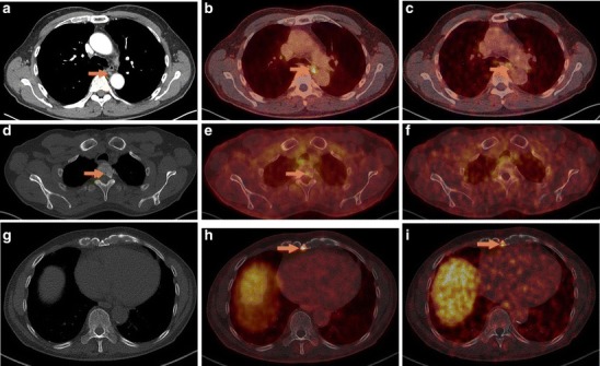
Procedures: In 14 selected patients with PSA relapse of prostate cancer, [(18)F]DCFPyL PET/X-ray computed tomography (CT) was performed in addition to [(68)Ga]Ga-PSMA-HBED-CC PET/CT. A systematic comparison was carried out between results obtained with both tracers with regard to the number of detected PSMA-positive lesions, the standardized uptake value (SUV)max and the lesion to background ratios.

Results: All suspicious lesions identified by [(68)Ga]Ga-PSMA-HBED-CC were also detected with [(18)F]DCFPyL. In three patients, additional lesions were observed using [(18)F]DCFPyL PET/CT. The mean SUVmax in the concordant [(18)F]DCFPyL PSMA-positive lesions was significantly higher as compared to [(68)Ga]Ga-PSMA-HBED-CC (14.5 vs. 12.2, p = 0.028, n = 15). The mean tumor to background ratios (n = 15) were significantly higher for [(18)F]DCFPyL compared to [(68)Ga]Ga-PSMA-HBED-CC using kidney, spleen, or parotid as reference organs (p = 0.006, p = 0.002, p = 0.008), but no significant differences were found using the liver (p = 0.167) or the mediastinum (p = 0.363) as reference organs.

Conclusion: [(18)F]DCFPyL PET/CT provided a high image quality and visualized small prostate lesions with excellent sensitivity. [(18)F]DCFPyL represents a highly promising alternative to [(68)Ga]Ga-PSMA-HBED-CC for PSMA-PET/CT imaging in relapsed prostate cancer.


Figures

Fig. 1
Fig. 1
Patient no. 12 with a rising PSA level of 3.87 ng/ml. In the past, the patient had retroperitoneal lymph node metastases, which were irradiated. On a Biograph mCT 128 scanner, comparison between a MIP (maximum intensity projection) with [18F]DCFPyL and b MIP with [68Ga]Ga-PSMA-HBED-CC. [18F]DCFPyL PET/CT clearly demonstrates several additional supradiaphragmatic PSMA-positive lesions. [68Ga]Ga-PSMA-HBED-CC PET/CT showed the supradiaphragmal lesion in the sternum.
Fig. 2
Fig. 2
Patient no. 12, Biograph mCT 128. [18F]DCFPyL PET/CT (b) was suspicious of an additional lymph node metastasis in the dorsal mediastinum, visible as a normal-sized lymph node in a low-dose CT scan without tracer accumulation on c [68Ga]Ga-PSMA-HBED-CC PET/CT. d Low-dose CT in bone window and e [18F]DCFPyL PET/CT were suspicious of a bone metastasis in vertebra Th3, barely detectable in f [68Ga]Ga-PSMA-HBED-CC PET/CT. h [18F]DCFPyL PET/CT was clearly suspicious of bone metastasis in the processus xiphoideus sterni, hard to detect in g low-dose CT or i [68Ga]Ga-PSMA-HBED-CC PET/CT.
Fig. 3
Fig. 3
Patient no. 2 with a rising PSA level of 4.7 ng/ml. a Low-dose CT in bone window and b [18F]DCFPyL PET/CT were suspicious of bone metastasis in vertebra L2, hard to discern in the c [68Ga]Ga-PSMA-HBED-CC PET/CT. Imaging was performed on a Biograph 16 PET/CT.
Fig. 4
Fig. 4
Patient no. 7 with a rising PSA level to 1.3 ng/ml. In the past, the patient had left iliac lymph node dissection with histologically confirmed lymph node metastases. Both tracers have shown a left iliac PSMA-avid lymph node, which was confirmed as metastasis (ac). Additionally, e the [18F]DCFPyL PET/CT was suspicious of a second PSMA-positive lymph node below the bifurcation of the v. cava inferior, visible as a normal-sized lymph node on d CT, hard to detected on f [68Ga]Ga-PSMA-HBED-CC PET/CT. a Low-dose CT scan, b [18F]DCFPyL PET/CT, and c [68Ga]Ga-PSMA-HBED-CC PET/CT at the level of the iliacae communes._d Low-dose CT scan, e [18F]DCFPyL PET/CT, and f [68Ga]Ga-PSMA-HBED-CC PET/CT at the level of the bifurcation. Imaging was performed on a Biograph 16 PET/CT.
Fig. 5
Fig. 5
a Ratio of SUVmax in lesions to SUVmean in the kidney and b ratio of SUVmax in lesions to SUVmean in the liver. The blue columns represent [68Ga]Ga-PSMA-HBED-CC PET/CT, and the red columns represent [18F]DCFPyL PET/CT. [18F]DCFPyL showed a higher tumor to background ratio than [68Ga]Ga-PSMA-HBED-CC when the kidney was used as a reference organ (p = 0.006, n = 15). The tumor to background ratio did not differ significantly between [18F]DCFPyL and [68Ga]Ga-PSMA-HBED-CC when the liver was used as a reference organ (p = 0.167, n = 15). LN lymph node metastasis, LR local relapse, BM bone metastasis.

References

    1. Silver DA, Pellicer I, Fair WR, et al. Prostate-specific membrane antigen expression in normal and malignant human tissues. Clin Cancer Res. 1997;3:81–85. - PubMed
    1. Afshar-Oromieh A, Malcher A, Eder M, et al. PET imaging with a [68Ga]gallium-labelled PSMA ligand for the diagnosis of prostate cancer: biodistribution in humans and first evaluation of tumour lesions. Eur J Nucl Med Mol Imaging. 2013;40:486–495. doi: 10.1007/s00259-012-2298-2. - DOI - PubMed
    1. Afshar-Oromieh A, Zechmann CM, Malcher A, et al. Comparison of PET imaging with a 68Ga-labelled PSMA ligand and 18F-choline-based PET/CT for the diagnosis of recurrent prostate cancer. Eur J Nucl Med Mol Imaging. 2014;41(1):11–20. doi: 10.1007/s00259-013-2525-5. - DOI - PMC - PubMed
    1. Afshar-Oromieh A, Haberkorn U, Schlemmer HP, et al. Comparison of PET/CT and PET/MRI hybrid systems using a 68Ga-labelled PSMA ligand for the diagnosis of recurrent prostate cancer: initial experience. Eur J Nucl Med Mol Imaging. 2014;41(5):887–897. doi: 10.1007/s00259-013-2660-z. - DOI - PubMed
    1. Afshar-Oromieh A, Avtzi E, Giesel FL, et al. The diagnostic value of PET/CT imaging with the 68Ga-labelled PSMA ligand HBED-CC in the diagnosis of recurrent prostate cancer. Eur J Nucl Med Mol Imaging. 2015;42:197–209. doi: 10.1007/s00259-014-2949-6. - DOI - PMC - PubMed

Publication types

MeSH terms