

Macroscopic optical physiological parameters correlate with microscopic proliferation and vessel area breast cancer signatures
- PMID: 26013572
- PMCID: PMC4487833
- DOI: 10.1186/s13058-015-0578-z
Macroscopic optical physiological parameters correlate with microscopic proliferation and vessel area breast cancer signatures
Abstract
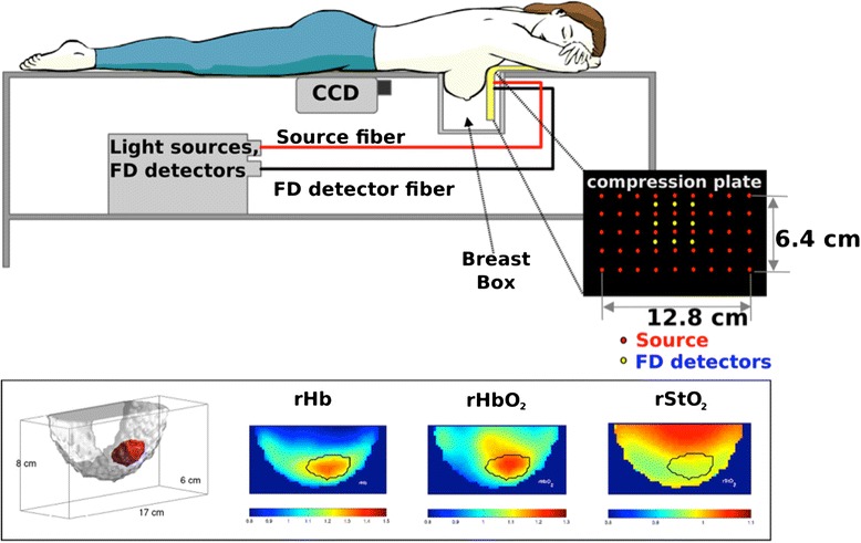
Introduction: Non-invasive diffuse optical tomography (DOT) and diffuse correlation spectroscopy (DCS) can detect and characterize breast cancer and predict tumor responses to neoadjuvant chemotherapy, even in patients with radiographically dense breasts. However, the relationship between measured optical parameters and pathological biomarker information needs to be further studied to connect information from optics to traditional clinical cancer biology. Thus we investigate how optically measured physiological parameters in malignant tumors such as oxy-, deoxy-hemoglobin concentration, tissue blood oxygenation, and metabolic rate of oxygen correlate with microscopic histopathological biomarkers from the same malignant tumors, e.g., Ki67 proliferation markers, CD34 stained vasculature markers and nuclear morphology.
Methods: In this pilot study, we investigate correlations of macroscopic physiological parameters of malignant tumors measured by diffuse optical technologies with microscopic histopathological biomarkers of the same tumors, i.e., the Ki67 proliferation marker, the CD34 stained vascular properties marker, and nuclear morphology.
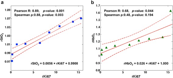
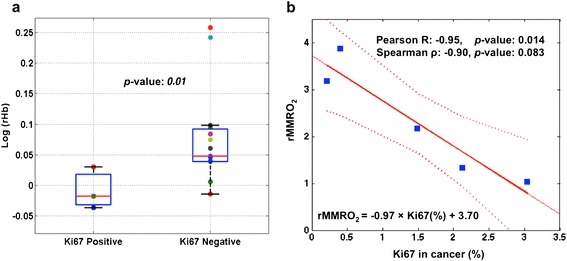
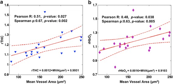
Results: The tumor-to-normal relative ratio of Ki67-positive nuclei is positively correlated with DOT-measured relative tissue blood oxygen saturation (R = 0.89, p-value: 0.001), and lower tumor-to-normal deoxy-hemoglobin concentration is associated with higher expression level of Ki67 nuclei (p-value: 0.01). In a subset of the Ki67-negative group (defined by the 15 % threshold), an inverse correlation between Ki67 expression level and mammary metabolic rate of oxygen was observed (R = -0.95, p-value: 0.014). Further, CD34 stained mean-vessel-area in tumor is positively correlated with tumor-to-normal total-hemoglobin and oxy-hemoglobin concentration. Finally, we find that cell nuclei tend to have more elongated shapes in less oxygenated DOT-measured environments.
Conclusions: Collectively, the pilot data are consistent with the notion that increased blood is supplied to breast cancers, and it also suggests that less conversion of oxy- to deoxy-hemoglobin occurs in more proliferative cancers. Overall, the observations corroborate expectations that macroscopic measurements of breast cancer physiology using DOT and DCS can reveal microscopic pathological properties of breast cancer and hold potential to complement pathological biomarker information.
Figures

Similar articles
-
US-localized diffuse optical tomography in breast cancer: comparison with pharmacokinetic parameters of DCE-MRI and with pathologic biomarkers.BMC Cancer. 2016 Feb 1;16:50. doi: 10.1186/s12885-016-2086-7. BMC Cancer. 2016. PMID: 26833069 Free PMC article.
-
Computational Model for Tumor Oxygenation Applied to Clinical Data on Breast Tumor Hemoglobin Concentrations Suggests Vascular Dilatation and Compression.PLoS One. 2016 Aug 22;11(8):e0161267. doi: 10.1371/journal.pone.0161267. eCollection 2016. PLoS One. 2016. PMID: 27547939 Free PMC article.
-
Parameters of dynamic contrast-enhanced MRI as imaging markers for angiogenesis and proliferation in human breast cancer.Med Sci Monit. 2015 Feb 1;21:376-82. doi: 10.12659/MSM.892534. Med Sci Monit. 2015. PMID: 25640082 Free PMC article.
-
Imaging in breast cancer: diffuse optics in breast cancer: detecting tumors in pre-menopausal women and monitoring neoadjuvant chemotherapy.Breast Cancer Res. 2005;7(6):279-85. doi: 10.1186/bcr1358. Epub 2005 Nov 28. Breast Cancer Res. 2005. PMID: 16457705 Free PMC article. Review.
-
The Role of Ki67 in Evaluating Neoadjuvant Endocrine Therapy of Hormone Receptor-Positive Breast Cancer.Front Endocrinol (Lausanne). 2021 Nov 3;12:687244. doi: 10.3389/fendo.2021.687244. eCollection 2021. Front Endocrinol (Lausanne). 2021. PMID: 34803903 Free PMC article. Review.
Cited by
-
Enhancement of diffuse correlation spectroscopy tissue blood flow measurement by acoustic radiation force.Biomed Opt Express. 2019 Dec 17;11(1):301-315. doi: 10.1364/BOE.381757. eCollection 2020 Jan 1. Biomed Opt Express. 2019. PMID: 32010518 Free PMC article.
-
Neoadjuvant chemotherapy for breast cancer: an evaluation of its efficacy and research progress.Front Oncol. 2023 Oct 3;13:1169010. doi: 10.3389/fonc.2023.1169010. eCollection 2023. Front Oncol. 2023. PMID: 37854685 Free PMC article. Review.
-
Diffuse Optical Characterization of the Healthy Human Thyroid Tissue and Two Pathological Case Studies.PLoS One. 2016 Jan 27;11(1):e0147851. doi: 10.1371/journal.pone.0147851. eCollection 2016. PLoS One. 2016. PMID: 26815533 Free PMC article.
-
Mechanical and hemodynamic responses of breast tissue under mammographic-like compression during functional dynamic optical imaging.Biomed Opt Express. 2020 Sep 3;11(10):5425-5441. doi: 10.1364/BOE.398110. eCollection 2020 Oct 1. Biomed Opt Express. 2020. PMID: 33149960 Free PMC article.
-
Diffuse Reflectance Spectroscopy of Changes in Tumor Microenvironment in Response to Different Doses of Radiation.Radiat Res. 2022 Dec 1;198(6):545-552. doi: 10.1667/RADE-21-00228.1. Radiat Res. 2022. PMID: 36240754 Free PMC article.
References
Publication types
MeSH terms
Substances
Grants and funding
LinkOut - more resources
Full Text Sources
Other Literature Sources
Medical
