. 2015 Jul 10;290(28):17349-66.
doi: 10.1074/jbc.M115.652305. Epub 2015 May 26.

Osteonecrosis of the Jaw Developed in Mice: DISEASE VARIANTS REGULATED BY γδ T CELLS IN ORAL MUCOSAL BARRIER IMMUNITY

Affiliations

Osteonecrosis of the Jaw Developed in Mice: DISEASE VARIANTS REGULATED BY γδ T CELLS IN ORAL MUCOSAL BARRIER IMMUNITY

Sil Park et al. J Biol Chem. .

Abstract

Osteonecrosis of the jaw (ONJ), an uncommon co-morbidity in patients treated with bisphosphonates (BP), occurs in the segment of jawbone interfacing oral mucosa. This study aimed to investigate a role of oral mucosal barrier γδ T cells in the pathogenesis of ONJ. Female C57Bl/6J (B6) mice received a bolus zoledronate intravenous injection (ZOL, 540 μg/kg), and their maxillary left first molars were extracted 1 week later. ZOL-treated mice (WT ZOL) delayed oral wound healing with patent open wounds 4 weeks after tooth extraction with characteristic oral epithelial hyperplasia. γδ T cells appeared within the tooth extraction site and hyperplastic epithelium in WT ZOL mice. In ZOL-treated γδ T cell null (Tcrd(-/-) ZOL) mice, the tooth extraction open wound progressively closed; however, histological ONJ-like lesions were identified in 75 and 60% of WT ZOL and Tcrd(-/-) ZOL mice, respectively. Although the bone exposure phenotype of ONJ was predominantly observed in WT ZOL mice, Tcrd(-/-) ZOL mice developed the pustule/fistula disease phenotype. We further addressed the role of γδ T cells from human peripheral blood (h-γδ T cells). When co-cultured with ZOL-pretreated human osteoclasts in vitro, h-γδ T cells exhibited rapid expansion and robust IFN-γ secretion. When h-γδ T cells were injected into ZOL-treated immunodeficient (Rag2(-/-) ZOL) mice, the oral epithelial hyperplasia developed. However, Rag2(-/-) ZOL mice did not develop osteonecrosis. The results indicate that γδ T cells are unlikely to influence the core osteonecrosis mechanism; however, they may serve as a critical modifier contributing to the different oral mucosal disease variations of ONJ.

Keywords: ONJ; T cell; bisphosphonate; mouse; mucosal immunology; osteonecrosis; pathogenesis; wound healing; γδ T cells.


Figures

FIGURE 1.
FIGURE 1.
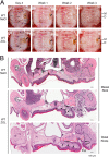
Tooth extraction wound healing in WT mice and the development of ONJ-like lesions in ZOL-treated WT mice (WT ZOL). A, time course of gingival/palatal tissue healing after maxillary first molar extraction (Ext). For control mice receiving 0.9% NaCl vehicle injection (WT NaCl), the open wound and surrounding tissue swelling (arrows) were progressively reduced from day 4 to week 2 after tooth extraction and healed by week 4. Mice administered a 540 μg/kg ZOL injection (WT ZOL) demonstrated slow wound healing, leaving the tooth extraction open wound (arrowheads) unclosed at the mesial root (M) with lesser degrees at the distal roots (D). The swelling of the gingival/palatal tissue (arrows) was restricted to the tooth extraction side. B, histological evaluations of tooth extraction wounds in WT NaCl and WT ZOL mice at week 4. Control WT NaCl mice exhibited complete wound healing as demonstrated by an extraction socket (black dotted line) filled with new bone formation and the re-establishment of oral epithelial integrity. The extraction wound was not closed in WT ZOL mice, enabling food and debris impaction. The jawbone exposure (Exp) was severe in the mesial root site; however, the distal root site revealed minimal but definitive jawbone exposure associated with abnormal epithelial growth resembling PEH (arrows). The tooth extraction socket (black dotted line) exhibited delayed bone formation. Osteonecrosis (red dotted line) was observed at the bone exposure site as well as the surface of the palatal/alveolar bone interfacing the oral mucosa with a localized dense inflammatory cell infiltration (arrowheads).
FIGURE 2.
FIGURE 2.
Appearance of γδ T cells in the oral mucosa after tooth extraction. A, palatal/gingival tissue of WT NaCl or WT ZOL mice was harvested 2 weeks after tooth extraction, and cells were dissociated by repeated enzymic digestions. Dissociated cells were incubated with CD45, CD3, γδTCR (GL3), or DX5 antibodies and examined by flow cytometry. In a representative analysis, CD45+ population contained CD3+ T cells at similar rates; however, the WT ZOL sample showed higher levels of GL3+ γδ T cells and DX5+ NK cells than the WT NaCl sample. B, to further identify γδ T cells, maxillary histological specimens were prepared from NaCl or ZOL-injected Tcrd-H2BEGFP mice 4 days or 2 weeks after tooth extraction (Ext.). Anti-GFP antibody was used to highlight GFP+ γδ T cells by immunohistology. A cluster of GFP+ γδ T cells was found in the connective tissue of the tooth extraction wound. C, number of GFP+ γδ T cells in the tooth extraction socket was found to be similar 4 days after tooth extraction in WT NaCl (n = 4) and WT ZOL (n = 4) mice. However, although in the control WT NaCl mice (n = 4), GFP+ γδ T cells decreased 2 weeks after tooth extraction, the GFP+ γδ T cell number increased in WT ZOL mice (n = 4). *, p < 0.05 D, in some WT ZOL mice, PEH-like oral epithelial hyperplasia (Epi Hyp) was observed at the tooth extraction site. GFP+ γδ T cells were abundant in the hyperplastic epithelium but not in normal oral epithelium.
FIGURE 3.
FIGURE 3.
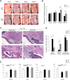
ONJ-like lesions in ZOL-treated γδ T cell null mice (Tcrd−/− ZOL). A, tooth extraction created the open wound (arrow) in the oral mucosa, which became visibly and consistently smaller in Tcrd−/− ZOL mice compared with WT ZOL mice at week 4. The swelling remained in the gingival/palatal tissues in Tcrd−/− ZOL mice (black dotted line). B, area of swelling normalized by the circumferential crown size of the contralateral first molar was significantly increased in Tcrd−/− ZOL mice compared with WT ZOL mice at weeks 2 and 4; *, p < 0.05. C, histological examination revealed that Tcrd−/− ZOL mice did not exhibit the bone exposure as observed in WT ZOL mice. The development of pustule (arrows; Pus) associated with epithelial fistulation (arrowheads) was demonstrated. The tooth extraction socket (black dotted line) healed uneventfully and was filled with woven bone. Osteonecrosis (red dotted line; Ost.Nec) was observed at the pustule areas and bone sequestration (red arrows). D, prevalence of necrotic jawbone exposure was significantly higher in WT ZOL mice than Tcrd−/− ZOL or WT NaCl control mice. However, Tcrd−/− mice exhibited more pustule/fistula formation than WT ZOL and WT NaCl mice. Both ONJ phenotypic variants were combined, and the prevalence of ONJ-like lesion development was found indistinguishable between WT ZOL and Tcrd−/− ZOL mice. *, p < 0.05; †, p = 0.06. E, micro-CT characterization of femur trabecular bone morphology of WT NaCl, WT ZOL, and Tcrd−/− ZOL mice. The effect of ZOL was shown as the increased bone mass and structure of trabecular bone of WT ZOL and Tcrd−/− ZOL mice. *, p < 0.05; **, p < 0.01.
FIGURE 4.
FIGURE 4.
Alveolar bone healing assessment by micro-CT imaging. A, WT NaCl control mice exhibited signs of osteolysis at week 2 as roughened alveolar and palatal bone surfaces (white arrowheads). Bone formation within the extraction socket (black arrowhead) appeared to complete the wound healing process at week 4. We noted that one WT NaCl mouse exhibited periodontal disease at the nonextraction side (white arrowhead). In WT ZOL and Tcrd−/− ZOL mice, the evidence of bone resorption was not clearly observed, and the extraction socket was not completely filled by new bone formation (black arrowheads). Severe periosteal reaction (black arrows) was observed at the molar extraction site in 53% of the WT ZOL mice. A periosteal reaction was also noted in one WT ZOL mouse at the contralateral nonextraction side with periodontal disease (confirmed with histology). Although the severity of periosteal reaction in Tcrd−/− ZOL mice was much attenuated (arrows), the sun-ray type of calcified spikes was observed both in WT ZOL and Tcrd−/− ZOL mice (SR with black arrow). B, micro-CT images were used to rate bone formation (0–2) and bone resorption (0–2) appearance. The bone-remodeling rate was expressed by combining the rates of bone formation and resorption in each animal. Active bone remodeling was suggested in WT NaCl control mice. Both WT ZOL and Tcrd−/− ZOL mice showed significantly decreased bone resorption. *, p < 0.05.
FIGURE 5.
FIGURE 5.
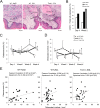
Characterization of osteonecrosis in WT NaCl, WT ZOL, and Tcrd−/− ZOL mice. A, localized inflammation (Inf) was evident near the surface of alveolar bone in WT NaCl mice. In WT ZOL mice, the intense inflammatory reaction was demonstrated in the oral mucosa (double-headed arrow; Inf). Abnormal epithelial tissue adhesion (Epi) was evident on the surface of partially necrotic alveolar bone (Ost. Nec.). In Tcrd−/− ZOL mice, inflammation was more diffused, and epithelial tissue did not reach the necrotic bone surface except at the pustule periphery. In both lesions, clusters of osteoclasts (arrowheads) were observed at and adjacent to the osteonecrosis site. B, CD45+ lymphocytes accounted for 30–45% of the dissociated cells from gingival/palatal tissue at day 4. The fraction of CD45+ cells decreased in WT NaCl mice at week 2. However, WT ZOL and Tcrd−/− ZOL mice exhibited similar fractions of CD45+ lymphocytes in the oral mucosa. C, area of osteonecrosis is presented as the percent of nonvital osteocytes over the total number of osteocytes on the oral side of palatal/alveolar bone. At week 4, the area of osteonecrosis was significantly larger in WT ZOL and Tcrd−/− ZOL mice than WT NaCl control mice. D, number of osteoclasts in the palatal/alveolar bone area suggested that the early increase in the tooth extraction wound of WT NaCl control mice followed by progressive decrease. Both WT ZOL and Tcrd−/− ZOL mice indicated a consistent appearance of osteoclasts throughout the experimental period, resulting in the elevated osteoclast number at week 4. *, p < 0.05; †, p = 0.06 compared with the WT NaCl control group. E, correlation between the osteonecrosis area and the number of OCs in week 1, week 2, and week 4 specimens. The WT NaCl group demonstrated the significant correlation between the osteonecrosis area and osteoclast number. By contrast, correlation pattern was similar in the WT ZOL and Tcrd−/− ZOL groups, which did not show statistical significance.
FIGURE 6.
FIGURE 6.
h-γδ T cell activation through co-culture with ZOL-pretreated hOCs and engraftment to Rag2−/− ZOL mice. A, positive selection of h-γδ T cells and h-CD3+ T cells from peripheral blood samples from healthy donors resulted in 76.9 and 96.4% purities, respectively. B, number of h-γδ T cells increased during the co-culture period of 6 days with ZOL-pretreated h-OC, whereas the number of h-CD3+ T cells decreased during the first 3 days. C, IFN-γ secreted by h-γδ T cells after the co-culture with h-OC was significantly higher than that of h-CD3+ T cells. *, p < 0.01. D, characterization of h-γδ T cells after the co-culture with ZOL-pretreated h-OC. h-γδ T cells were positive for CD3 and negative for CD19, indicating their T cell signature. Over 80% of h-γδ T cells showed CD69-positive, suggesting lymphocyte activation after co-culturing with ZOL-pretreated h-OC. E, representative profile of mouse immune effector cells in bone marrow of Rag2−/− ZOL mice engrafted with activated h-γδ T cells. The deficiency of mouse CD45+ CD19+ B cells and mouse CD45+ γδTCR (GL3)+ γδ T cells was indicated. Mouse CD45+ cells contained F4/80+ monocytes. Human cells appeared to be h-CD45+ h-γδTCR+ or h-CD3+ h-γδTCR+ suggesting the successful engraftment of h-γδ T cells.
FIGURE 7.
FIGURE 7.
ONJ lesions of Rag2−/− mice and the effect of h-γδ T cell engraftment. A, accelerated tooth extraction wound healing was observed in Rag2−/− mice. Two weeks after first molar extraction, both Rag2−/− NaCl and Rag2−/− ZOL mice exhibited uneventful healing (arrows) without oral mucosa swelling. Tooth extraction wound was completely closed in all Rag2−/− NaCl mice (n = 5), whereas Rag2−/− ZOL mice (n = 5) showed nonkeratinized oral mucosa covering the extraction socket or small open wound with food and debris impaction. Histologically, the extraction socket (Soc) was filled with newly formed bone in both Rag2−/− NaCl and Rag2−/− ZOL mice. The area of osteonecrosis (Ost Nec) was small. There were small food and debris impactions (arrow), which appeared to be integrated in the healed tissue. B, Rag2−/− ZOL mice engrafted with h-γδ T cells (n = 4) similarly showed the wound closure without swelling; however, there were unusual pits and irregular papilloma-like tissues (arrows) were present at the first molar extraction site. Histologically, all Rag2−/− ZOL h-γδ T cell mice exhibited various degrees of oral epithelial hyperplasia (Epi, arrows). In some specimens, epithelial “pouch” was found in the deep connective tissue, which often, but not always, contained small food and debris. The extraction socket (Soc) containing the epithelial pouch showed delayed bone formation. Osteonecrosis was found in small areas, associated with osteoclasts (arrowheads). C, osteonecrosis was determined by the bone area of containing ≥5 empty osteocyte lacunae over the total alveolar bone area. Because all Rag2−/− mice were housed with autoclaved cellulose-based bedding, a separate set of WT NaCl and WT ZOL mice (n = 5 in each group) was housed in the same environment after tooth extraction. Tooth extraction wound healing appeared to be more favorable in this environment; however, WT ZOL mice exhibited open tooth extraction wounds with significantly larger osteonecrosis areas. The osteonecrosis area of Rag2−/− ZOL mice remained small at the levels of WT NaCl and Rag2−/− NaCl mice. The engraftment of h-γδ T cells to Rag2−/− ZOL mice did not increase the osteonecrosis area. *, p < 0.05 compared with the WT NaCl control group.
FIGURE 8.
FIGURE 8.
Osteoclast behavior in Rag2−/− mice with h-γδ T cell engraftment. A, tartrate-resistant acid phosphatase staining revealed osteoclasts on the surface of palatal bone (arrows) interfacing the area of oral mucosa inflammation (Inf) in WT ZOL mice. Strikingly, osteoclasts on the palatal surface were nearly absent in Rag2−/− ZOL mice, whereas osteoclasts appeared in the bone remodeling area of tooth extraction socket (Soc). The number of osteoclasts normalized by the bone surface was significantly reduced on the palatal bone surface in Rag2−/− regardless of ZOL treatment as well as h-γδ T cell engraftment. The osteoclast number in the extraction socket was normalized in Rag2−/− NaCl and Rag2−/− ZOL mice, whereas h-γδ T cell-engrafted mice remained high. *, p < 0.05 compared with the WT NaCl control group. B, stimulatory effect of m-OC on h-γδ T cells and m-γδ T cells was assessed in vitro. ELISA revealed that m-OC with ZOL pretreatment specifically activated the IFN-γ secretion by h-γδ T cells but not by m-γδ T cell. However, nonspecific activation by m-OC was observed in both h-γδ T cells and m-γδ T cells, which secreted higher levels of IFN-γ. *, p < 0.05 compared with the h-γδ T cell or m-γδ T cell alone group at each time point.

References

    1. Reid I. R., Cornish J. (2012) Epidemiology and pathogenesis of osteonecrosis of the jaw. Nat. Rev. Rheumatol. 8, 90–96 - PubMed
    1. Saad F., Brown J. E., Van Poznak C., Ibrahim T., Stemmer S. M., Stopeck A. T., Diel I. J., Takahashi S., Shore N., Henry D. H., Barrios C. H., Facon T., Senecal F., Fizazi K., Zhou L., et al. (2012) Incidence, risk factors, and outcomes of osteonecrosis of the jaw: integrated analysis from three blinded active-controlled phase III trials in cancer patients with bone metastases. Ann. Oncol. 23, 1341–1347 - PubMed
    1. Khosla S., Burr D., Cauley J., Dempster D. W., Ebeling P. R., Felsenberg D., Gagel R. F., Gilsanz V., Guise T., Koka S., McCauley L. K., McGowan J., McKee M. D., Mohla S., Pendrys D. G., et al. (2007) Bisphosphonate-associated osteonecrosis of the jaw: report of a task force of the American Society for Bone and Mineral Research. J. Bone Miner. Res. 22, 1479–1491 - PubMed
    1. Ruggiero S. L., Dodson T. B., Assael L. A., Landesberg R., Marx R. E., Mehrotra B. (2009) American Association of Oral and Maxillofacial Surgeons position paper on bisphosphonate-related osteonecrosis of the jaws–2009 update. J. Oral Maxillofac. Surg. 67, 2–12 - PubMed
    1. Cetiner S., Sucak G. T., Kahraman S. A., Aki S. Z., Kocakahyaoglu B., Gultekin S. E., Cetiner M., Haznedar R. (2009) Osteonecrosis of the jaw in patients with multiple myeloma treated with zoledronic acid. J. Bone Miner. Metab. 27, 435–443 - PubMed

Publication types

MeSH terms