

Contrast Ultrasound Imaging of the Aorta Does Not Affect Progression of Atherosclerosis or Cardiovascular Biomarkers in ApoE-/- Mice
- PMID: 26014332
- PMCID: PMC4471945
- DOI: 10.7863/ultra.34.6.1115
Contrast Ultrasound Imaging of the Aorta Does Not Affect Progression of Atherosclerosis or Cardiovascular Biomarkers in ApoE-/- Mice
Abstract
Objectives: Ultrasound contrast agents (UCAs) enhance cardiovascular ultrasound imaging. Adverse biological effects have occurred after administration of UCAs, and more research is needed for a comprehensive understanding of the risks involved. We used the ApoE(-/-) mouse model of atherosclerosis to characterize the effects of ultrasound and UCAs on atherosclerosis and plasma biomarkers.
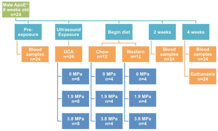
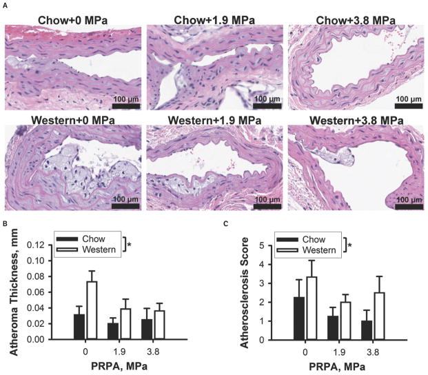
Methods: Male ApoE(-/-) mice (8 weeks old; n = 24) were intravenously infused with a UCA (2 × 10(10) Definity microbubbles per hour; Lantheus Medical Imaging, North Billerica, MA) and exposed to 2.8-MHz center frequency ultrasound (10 Hz pulse repetition frequency, 1.4 microseconds pulse duration, 2 minutes exposure duration, and 2 sites) at 1 of 3 derated peak rarefactional pressure amplitudes (0, 1.9, or 3.8 MPa), and then consumed either a chow or Western diet for 4 weeks (n = 4 per group). Blood plasma samples were collected before ultrasound exposure and at 2 and 4 weeks after exposure and assayed for total cholesterol and von Willebrand Factor (vWF). A pathologist measured atheroma thickness in formalin-fixed, hematoxylin-eosin-stained transverse aorta sections and scored them for severity of atherosclerosis.
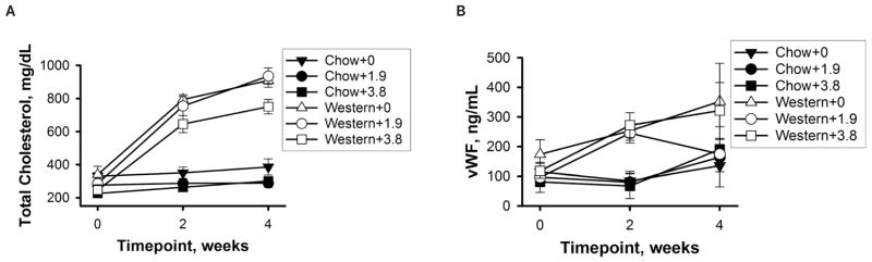
Results: Plasma total cholesterol initially averaged 286 mg/dL in the Western diet group and increased to 861 mg/dL after 4 weeks on the diet (P < .0001). Total cholesterol did not increase significantly in the chow diet group. Plasma vWF increased after 2 weeks on the Western diet (P < .0001). Atheroma thickness was greater in animals consuming the Western diet than in chow-fed animals (P < .05). Ultrasound had no significant effect on plasma total cholesterol, plasma vWF, or atheroma thickness.
Conclusions: Contrast ultrasound did not increase the severity of atherosclerosis or alter cardiovascular biomarkers in the ApoE(-/-) mouse model.
Keywords: atherosclerosis; biomarkers; cardiovascular disease; contrast agents; endothelium; microbubbles; ultrasound.
© 2015 by the American Institute of Ultrasound in Medicine.
Figures
Similar articles
-
Contrast Ultrasound Imaging Does Not Affect Heat Shock Protein 70 Expression in Cholesterol-Fed Rabbit Aorta.J Ultrasound Med. 2015 Jul;34(7):1209-16. doi: 10.7863/ultra.34.7.1209. J Ultrasound Med. 2015. PMID: 26112623 Free PMC article.
-
Contrast ultrasound imaging of the aorta alters vascular morphology and circulating von Willebrand factor in hypercholesterolemic rabbits.J Ultrasound Med. 2012 May;31(5):711-20. doi: 10.7863/jum.2012.31.5.711. J Ultrasound Med. 2012. PMID: 22535718 Free PMC article.
-
Monitoring inflammation injuries in the progression of atherosclerosis with contrast enhanced ultrasound molecular imaging.PLoS One. 2017 Oct 5;12(10):e0186155. doi: 10.1371/journal.pone.0186155. eCollection 2017. PLoS One. 2017. PMID: 28982198 Free PMC article.
-
Contrast-enhanced ultrasound: clinical applications in patients with atherosclerosis.Int J Cardiovasc Imaging. 2016 Jan;32(1):35-48. doi: 10.1007/s10554-015-0713-z. Epub 2015 Jul 24. Int J Cardiovasc Imaging. 2016. PMID: 26206524 Free PMC article. Review.
-
Ultrasound imaging for risk assessment in atherosclerosis.Int J Mol Sci. 2015 Apr 29;16(5):9749-69. doi: 10.3390/ijms16059749. Int J Mol Sci. 2015. PMID: 25938969 Free PMC article. Review.
Cited by
-
Preclinical techniques to investigate exercise training in vascular pathophysiology.Am J Physiol Heart Circ Physiol. 2021 Apr 1;320(4):H1566-H1600. doi: 10.1152/ajpheart.00719.2020. Epub 2021 Jan 1. Am J Physiol Heart Circ Physiol. 2021. PMID: 33385323 Free PMC article.
References
-
- Senior R, Shah BN. Myocardial contrast echocardiography for simultaneous assessment of function and perfusion in real time: a technique comes of age. Circulation. 2012;126:1182–1184. - PubMed
-
- Porter T, Li S, Kilzer K, Deligonul U. Correlation between quantitative angiographic lesion severity and myocardial contrast intensity during a continuous infusion of perfluorocarbon-containing microbubbles. J Am Soc Echocardiogr. 1998;11:702–710. - PubMed
-
- Akkus Z, Hoogi A, Renaud G, et al. New quantification methods for carotid intra-plaque neovascularization using contrast-enhanced ultrasound. Ultrasound Med Biol. 2014;40:25–36. - PubMed
-
- Kitzman DW, Goldman ME, Gillam LD, Cohen JL, Aurigemma GP, Gottdiener JS. Efficacy and safety of the novel ultrasound contrast agent perflutren (Definity) in patients with suboptimal baseline left ventricular echocardiographic images. Am J Cardiol. 2000;86:669–674. - PubMed
-
- Reilly JP, Tunick PA, Timmermans RJ, Stein B, Rosenzweig BP, Kronzon I. Contrast echocardiography clarifies uninterpretable wall motion in intensive care unit patients. J Am Coll Cardiol. 2000;35:485–490. - PubMed
Publication types
MeSH terms
Substances
Grants and funding
LinkOut - more resources
Full Text Sources
Other Literature Sources
Medical
Miscellaneous
