. 2015 May 27;16(1):407.
doi: 10.1186/s12864-015-1568-3.

Transcriptome and venom proteome of the box jellyfish Chironex fleckeri

Affiliations

Transcriptome and venom proteome of the box jellyfish Chironex fleckeri

Diane L Brinkman et al. BMC Genomics. .

Abstract

Background: The box jellyfish, Chironex fleckeri, is the largest and most dangerous cubozoan jellyfish to humans. It produces potent and rapid-acting venom and its sting causes severe localized and systemic effects that are potentially life-threatening. In this study, a combined transcriptomic and proteomic approach was used to identify C. fleckeri proteins that elicit toxic effects in envenoming.

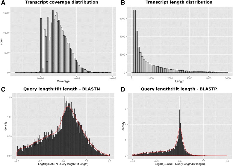
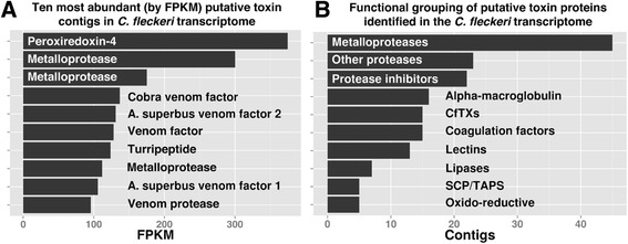
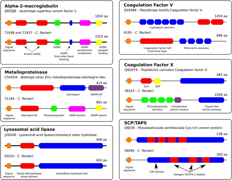
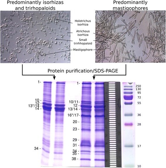
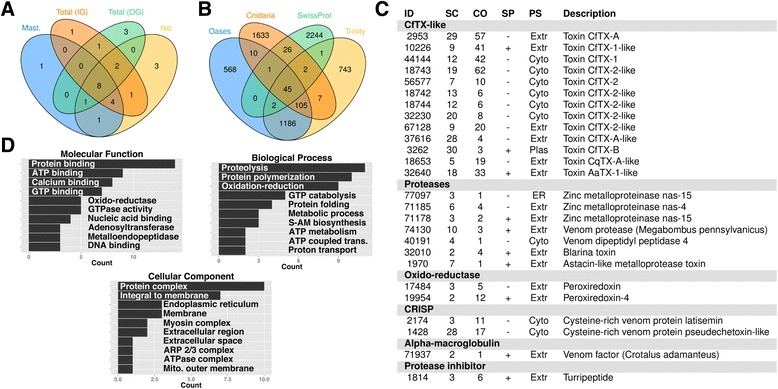
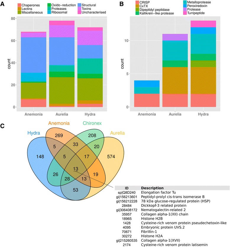
Results: More than 40,000,000 Illumina reads were used to de novo assemble ∼ 34,000 contiguous cDNA sequences and ∼ 20,000 proteins were predicted based on homology searches, protein motifs, gene ontology and biological pathway mapping. More than 170 potential toxin proteins were identified from the transcriptome on the basis of homology to known toxins in publicly available sequence databases. MS/MS analysis of C. fleckeri venom identified over 250 proteins, including a subset of the toxins predicted from analysis of the transcriptome. Potential toxins identified using MS/MS included metalloproteinases, an alpha-macroglobulin domain containing protein, two CRISP proteins and a turripeptide-like protease inhibitor. Nine novel examples of a taxonomically restricted family of potent cnidarian pore-forming toxins were also identified. Members of this toxin family are potently haemolytic and cause pain, inflammation, dermonecrosis, cardiovascular collapse and death in experimental animals, suggesting that these toxins are responsible for many of the symptoms of C. fleckeri envenomation.

Conclusions: This study provides the first overview of a box jellyfish transcriptome which, coupled with venom proteomics data, enhances our current understanding of box jellyfish venom composition and the molecular structure and function of cnidarian toxins. The generated data represent a useful resource to guide future comparative studies, novel protein/peptide discovery and the development of more effective treatments for jellyfish stings in humans. (Length: 300).


Figures

Figure 1
Figure 1
Summary of C. fleckeri assembly. A. The coverage of assembled transcripts after mapping of raw sequences back to the assembly using RSEM; B. The transcript length distribution; C. The distribution of the ratio of BLAST query length to BLAST hit length for transcripts when searched against a H. magnipapillata EST database using blastn. The EST database was obtained from Metazome (http://www.metazome.net/) and generated as part of the H. magnipapillata genome project; and D. Distribution of BLAST query length to hit length ratios for C. fleckeri predicted proteins searched against the SwissProt database using blastp.
Figure 2
Figure 2
Potential toxin encoding transcripts identified in the transcriptome of C. fleckeri. A. The ten most abundant (by FPKM) transcripts encoding potential toxin proteins; B. Number of different transcripts in the assembly encoding potential toxin proteins from the ten toxin families identified in the transcriptome. In both A. and B. potential toxins were identified after screening against the SwissProt animal toxin database.
Figure 3
Figure 3
Domain structure of C. fleckeri transcripts encoding potential toxin proteins. Using InterProScan, the domain structure of potential C. fleckeri toxins were compared to their respective top scoring BLAST hit from the SwissProt animal toxin database. In three cases the domain structure of potential toxins was the same as those identified from the database but in the other cases different domain structures suggest functional divergence.
Figure 4
Figure 4
Light microscopy of nematocysts used in proteomic analysis and SDS-PAGE gels of venom proteins. Two nematocyst preparations were analyzed; a preparation containing predominantly mastigophores (right) and a preparation enriched in isorhizas and trirhopaloids (left). Different morphological types are indicated (magnification 200x). Extracts of the nematocyst preparations were fractionated using SDS-PAGE (bottom) and proteins identified using tandem mass spectrometry. Forty-one gel slices were excised from each lane, as indicated by the aligned metal grid (right). Selected gel slices corresponding to major protein bands are indicated on the gel. Where a protein band was divided between two gel slices, an asterisk denotes the gel slice containing the majority of that protein.
Figure 5
Figure 5
MS/MS analysis of C. fleckeri venom. A. Venn diagram showing the numbers of potential toxin proteins identified in each MS/MS experiment. Abbreviations used, Mast. — nematocyst sample containing predominantly mastigophores; Iso. — nematocyst sample containing predominantly isorhizas and trirhopaloids; Total (IG) — total nematocyst sample fractionated using SDS-PAGE; and Total (OG) — total nematocyst sample fractionated using peptide OFFGEL electrophoresis; B. Venn diagram showing overlap in significant peptide identifications in three additional databases searches using MS/MS data. The databases depicted are 1.) Oases — predicted protein dataset from Oases assembly; 2.) Trinity — predicted dataset Trinity assembly; 3.) Cnidaria — all cnidarian proteins from the GenBank non-redundant protein database; and 4.) SwissProt — the Uniprot SwissProt database; C. Proteins identified in the venom using MS/MS that had been previously identified as potential toxins during the analysis of the transcriptome; D. GO terms associated with proteins identified in the venom of C. fleckeri using MS/MS.
Figure 6
Figure 6
Comparison of cnidarian venom proteomes. A. Proteins from the C. fleckeri proteome with corresponding proteins in H. magnipapillata, A. aurita and A. viridis by functional category; B. Potential toxins from C. fleckeri with corresponding proteins in the same species; C. Venn diagram depicting the overlap in proteins identified in the proteomes of four cnidarian species.
Figure 7
Figure 7
Phylogenetic tree of characterized CfTX toxin proteins. Phylogenetic tree depicting the grouping of CfTX-like proteins in cnidaria. Proteins names and accessions are shown. Proteins identified in this study are depicted with an asterisk and those from [27] with a double asterisk. The tree was produced using MUSCLE and PhyML for tree building and the aLRT statistical test [55] was used for branch support.

References

    1. Brinkman DL, Burnell JN. Biochemical and molecular characterisation of cubozoan protein toxins. Toxicon. 2009;54:1162–73. doi: 10.1016/j.toxicon.2009.02.006. - DOI - PubMed
    1. Brinkman DL, Konstantakopoulos N, McInerney BV, Mulvenna J, Seymour JE, Isbister GK, et al. Chironex fleckeri (box jellyfish) venom proteins: Expansion of a cnidarian toxin family that elicits variable cytolytic and cardiovascular effects. J Biol Chem. 2014;289:4798–812. doi: 10.1074/jbc.M113.534149. - DOI - PMC - PubMed
    1. Chaousis S, Smout M, Wilson D, Loukas A, Mulvenna J, Seymour J. Rapid short term and gradual permanent cardiotoxic effects of vertebrate toxins from Chironex fleckeri (Australian box jellyfish) venom. Toxicon. 2014;80:17–26. doi: 10.1016/j.toxicon.2014.01.007. - DOI - PubMed
    1. Hughes RJ, Angus JA, Winkel KD, Wright CE. A pharmacological investigation of the venom extract of the australian box jellyfish, Chironex fleckeri, in cardiac and vascular tissues. Toxicol Lett. 2012;209:11–20. doi: 10.1016/j.toxlet.2011.11.025. - DOI - PubMed
    1. Yanagihara AA, Shohet RV. Cubozoan venom-induced cardiovascular collapse is caused by hyperkalemia and prevented by zinc gluconate in mice. PLoS ONE. 2012;7:e51368. doi: 10.1371/journal.pone.0051368. - DOI - PMC - PubMed

Publication types

Substances

LinkOut - more resources