

A Highly Expressed Human Protein, Apolipoprotein B-100, Serves as an Autoantigen in a Subgroup of Patients With Lyme Disease
- PMID: 26014802
- PMCID: PMC4633766
- DOI: 10.1093/infdis/jiv310
A Highly Expressed Human Protein, Apolipoprotein B-100, Serves as an Autoantigen in a Subgroup of Patients With Lyme Disease
Abstract
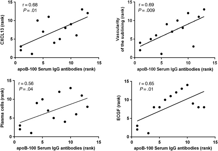
To discover novel autoantigens associated with Lyme arthritis (LA), we identified T-cell epitopes presented in vivo by human leukocyte antigen (HLA)-DR molecules in patients' inflamed synovial tissue or joint fluid and tested each epitope for autoreactivity. Using this approach, we identified the highly expressed human protein, apolipoprotein B-100 (apoB-100), as a target of T- and B-cell responses in a subgroup of LA patients. Additionally, the joint fluid of these patients had markedly elevated levels of apoB-100 protein, which may contribute to its autoantigenicity. In patients with antibiotic-refractory LA, the magnitude of apoB-100 antibody responses correlated with increased numbers of plasma cells in synovial tissue, greater numbers and activation of endothelial cells, and more synovial fibroblast proliferation. Thus, a subset of LA patients have high levels of apoB-100 in their joints and autoreactive T- and B-cell responses to the protein, which likely contributes to pathogenic autoimmunity in patients with antibiotic-refractory LA.
Keywords: Borrelia burgdorferi; Lyme arthritis; Lyme disease; apolipoprotein B; autoantibodies; autoimmunity; erythema migrans.
© The Author 2015. Published by Oxford University Press on behalf of the Infectious Diseases Society of America. All rights reserved. For Permissions, please e-mail: journals.permissions@oup.com.
Figures


Similar articles
-
Annexin A2 is a target of autoimmune T and B cell responses associated with synovial fibroblast proliferation in patients with antibiotic-refractory Lyme arthritis.Clin Immunol. 2015 Oct;160(2):336-41. doi: 10.1016/j.clim.2015.07.005. Epub 2015 Jul 15. Clin Immunol. 2015. PMID: 26187145 Free PMC article.
-
Matrix metalloproteinase-10 is a target of T and B cell responses that correlate with synovial pathology in patients with antibiotic-refractory Lyme arthritis.J Autoimmun. 2016 May;69:24-37. doi: 10.1016/j.jaut.2016.02.005. Epub 2016 Feb 26. J Autoimmun. 2016. PMID: 26922382 Free PMC article.
-
Identification of Major Histocompatibility Complex Class II Epitopes From Lyme Autoantigen Apolipoprotein B-100 and Borrelia burgdorferi Mcp4 in Murine Lyme Arthritis.J Infect Dis. 2024 Aug 14;230(Supplement_1):S27-S39. doi: 10.1093/infdis/jiae324. J Infect Dis. 2024. PMID: 39140726 Free PMC article.
-
Autoimmune mechanisms in antibiotic treatment-resistant lyme arthritis.J Autoimmun. 2001 May;16(3):263-8. doi: 10.1006/jaut.2000.0495. J Autoimmun. 2001. PMID: 11334491 Review.
-
Molecular mimicry to Borrelia burgdorferi: pathway to autoimmunity?Autoimmunity. 2004 Aug;37(5):387-92. doi: 10.1080/08916930410001713098. Autoimmunity. 2004. PMID: 15621562 Review.
Cited by
-
Annexin A2 is a target of autoimmune T and B cell responses associated with synovial fibroblast proliferation in patients with antibiotic-refractory Lyme arthritis.Clin Immunol. 2015 Oct;160(2):336-41. doi: 10.1016/j.clim.2015.07.005. Epub 2015 Jul 15. Clin Immunol. 2015. PMID: 26187145 Free PMC article.
-
CD1b presents self and Borrelia burgdorferi diacylglycerols to human T cells.Eur J Immunol. 2019 May;49(5):737-746. doi: 10.1002/eji.201847949. Epub 2019 Mar 20. Eur J Immunol. 2019. PMID: 30854633 Free PMC article.
-
Borrelia burgdorferi peptidoglycan is a persistent antigen in patients with Lyme arthritis.Proc Natl Acad Sci U S A. 2019 Jul 2;116(27):13498-13507. doi: 10.1073/pnas.1904170116. Epub 2019 Jun 17. Proc Natl Acad Sci U S A. 2019. PMID: 31209025 Free PMC article.
-
Two rheumatoid arthritis-specific autoantigens correlate microbial immunity with autoimmune responses in joints.J Clin Invest. 2017 Aug 1;127(8):2946-2956. doi: 10.1172/JCI93450. Epub 2017 Jun 26. J Clin Invest. 2017. PMID: 28650341 Free PMC article.
-
Cell-Mediated Cytotoxicity in Lyme Arthritis.Arthritis Rheumatol. 2023 May;75(5):782-793. doi: 10.1002/art.42408. Epub 2023 Mar 20. Arthritis Rheumatol. 2023. PMID: 36413215 Free PMC article.
References
-
- Steere AC, Grodzicki RL, Kornblatt AN et al. . The spirochetal etiology of Lyme disease. N Engl J Med 1983; 308:733–40. - PubMed
-
- Benach JL, Bosler EM, Hanrahan JP et al. . Spirochetes isolated from the blood of two patients with Lyme disease. N Engl J Med 1983; 308:740–2. - PubMed
-
- Steere AC. Lyme disease. N Engl J Med 2001; 345:115–25. - PubMed
-
- Steere AC, Angelis SM. Therapy for Lyme arthritis: strategies for the treatment of antibiotic-refractory arthritis. Arthritis Rheum 2006; 54:3079–86. - PubMed
Publication types
MeSH terms
Substances
Grants and funding
LinkOut - more resources
Full Text Sources
Other Literature Sources
Medical
Research Materials
Miscellaneous
