. 2015 May 27:5:10283.
doi: 10.1038/srep10283.

Centrin-2 (Cetn2) mediated regulation of FGF/FGFR gene expression in Xenopus

Affiliations

Centrin-2 (Cetn2) mediated regulation of FGF/FGFR gene expression in Xenopus

Jianli Shi et al. Sci Rep. .

Abstract

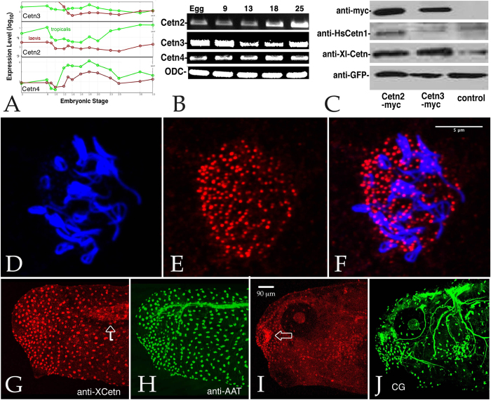
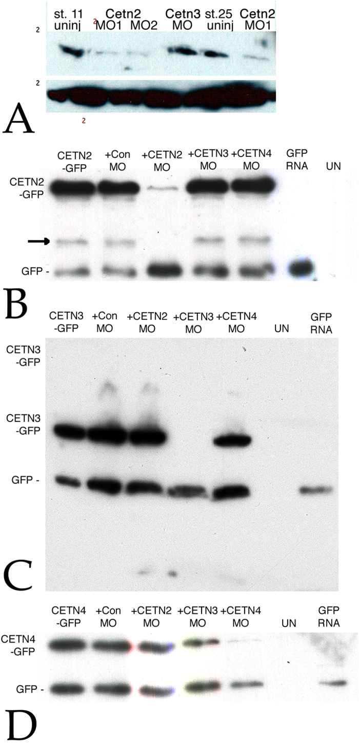
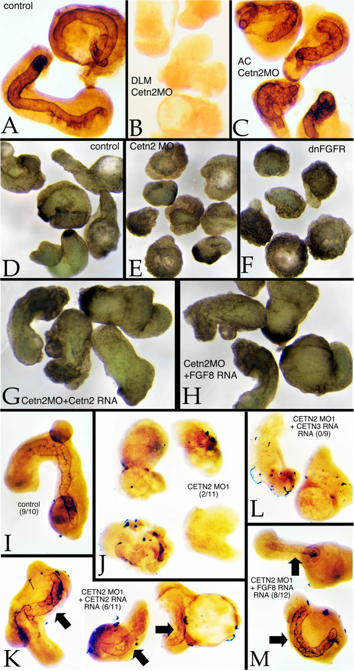
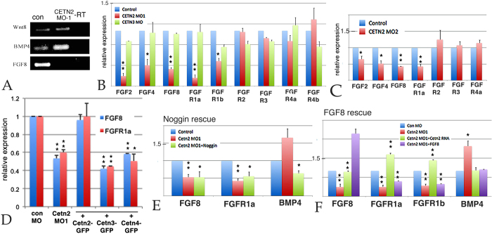
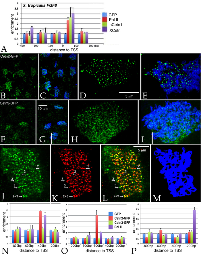
Centrins (Cetns) are highly conserved, widely expressed, and multifunctional Ca(2+)-binding eukaryotic signature proteins best known for their roles in ciliogenesis and as critical components of the global genome nucleotide excision repair system. Two distinct Cetn subtypes, Cetn2-like and Cetn3-like, have been recognized and implicated in a range of cellular processes. In the course of morpholino-based loss of function studies in Xenopus laevis, we have identified a previously unreported Cetn2-specific function, namely in fibroblast growth factor (FGF) mediated signaling, specifically through the regulation of FGF and FGF receptor RNA levels. Cetn2 was found associated with the RNA polymerase II binding sites of the Cetn2-regulated FGF8 and FGFR1a genes, but not at the promoter of a gene (BMP4) whose expression was altered indirectly in Cent2 morphant embryos. These observations point to a previously unexpected role of Cetn2 in the regulation of gene expression and embryonic development.


Figures

Figure 1
Figure 1
A: All three Cetn RNAs are present throughout the course of early development (data derived from Yanai et al. (2011)). B: This result was confirmed by RT-PCR analyses of Cetn2a, Cetn3l, and Cetn4 RNAs using ornithine decarboxylase (ODC) as a normalization control (embryonic stages are noted). C: Embryos injected with RNA (200 pg) encoding GFP alone or together with Cetn2a-myc or Cetn3l-myc were harvested at stage 11 and analyzed by SDS-PAGE-immunoblot. The anti-human Cetn1 antibody reacted preferentially with X. laevis Cetn2, while the anti-XlCetn antibody reacted with Cetn2 and Cetn3, as well as Cetn4 (data not shown). Ectodermal explants were fixed when sibling control embryos reached stage 18 and stained with anti-acetylated α-tubulin (AAT)(D) and anti-XlCetn antibodies (E; F displays the overlap of images in parts D and E); this revealed the localization of Cetns to the basal body region of cilia. A similar analysis was carried out on whole embryos (G,H - stage 25, I,J-stage 35) stained with anti-XlCetn (G,I) and anti-acetylated α-tubulin (H,J). Anti-Cetn staining of the myotome (arrow in part G) and Cetn’s localization to the olfactory region of the later stage embryo (arrow in part I) was obvious, as was its absence from the cement gland (“CG” in part J). Scale bar in part F marks 5 μm in parts D-F, scale bar in part I marks 90 μm in parts G-J.
Figure 2
Figure 2
A: Embryos were injected into both cells at the two cell stage with RNAs encoding GFP (150 pgs per embryo) either alone or together with Cetn2MO1, Cetn2MO2, or Cetn3MO (10 ngs/side, 20 ngs total per embryo); at stage 11 or 25 the embryos were analyzed by SDS-PAGE and immunoblot using the anti-Human Cetn-1 antibody (which reacts preferentially with Cetn2 compared to Cetn3. There was a clear decrease in Cetn2 protein levels, persisting through stage 25. To confirm the specificities of the Cetn MOs both blastomeres of two cell embryos were injected with RNAs encoding GFP (200 pg/side) and RNAs encoding Cetn2a-GFP (B), Cetn3l-GFP (C), or Cetn4-GFP (D) RNAs with (“+”) or without Cetn MO (10 ng/side). These Cetn RNAs contain the target sequence of the corresponding morpholino. In addition, uninjected (“UN”) and embryos injected with GFP RNA alone were examined as controls for antibody specificity. Injected embryos were harvested at stage 11. Immunoblot analyses were carried out using an anti-rabbit GFP antibody. An apparent breakdown product of the Cetn2-GFP construct is indicated by the arrow in the Cetn2 MO panel.
Figure 3
Figure 3
Animal cap/dorsal axial mesodermal zone (AC/DAMZ) explants were prepared from experimentally manipulated embryos when control (intact) embryos had reached stage 25. In wild type explants (A) staining with the anti-keratan sulfate antibody MZ15 revealed explant elongation and notochord formation. Both were absent in wild type AC/Cetn2 morphant DAMZ explants (B). Notochord formation occurred in Cetn2 morphant AC/wild type DAMZ explants (C). A comparison of dorsal axial mesoderm explant morphology (D-H) revealed the elongation of control explants (D), this elongation phenotype was absent in Cetn2 morphant explants (E) and dominant-negative FGFR RNA injected explants (F). In Cetn2 morphant explants, the elongation phenotype was rescued by either Cetn2-GFP (G) or FGF8 (H) RNA injection (200 pgs/embryo). Morpholinos were injected at 10 ng/embryo. Staining with MZ15 revealed the presence of notochordal tissues in control (I) explants, its absence in Cetn2 morphant explants (J), and its reappearance in Cetn2 RNA (K) and FGF8 (M), but not in Cetn3 RNA (L) injected Cetn2 morphant explants - number of explants with notochord staining per total number of explants is presented in brackets in panels I-M.
Figure 4
Figure 4
A: PCR analysis of control and Cetn2 MO embryos; there was a clear increase in BMP4 RNA, the disappearance of FGF8 RNA, and no apparent effect on Wnt8 RNA: B: qPCR analysis of Control, Cetn2MO1 and Cetn3MO embryos, injected in both cells of a two cell embryo and harvested at stage 11. C: A similar analysis carried out with the Cetn2MO2. D: qPCR analysis of embryos injected in both cells of a two cell embryo with Cetn2MO1 alone or together with Cetn2-GFP, Cetn3-GFP, or Cetn4-GFP RNAs (RNAs injected at 200 pg/side, total 400 pg/embryo; MOs injected at 10 ng/side, total 20 ng/embryo). While Cetn2-GFP RNA rescued the morphant phenotype Cetn3 or Cetn4 RNAs did not. E: qPCR analysis of embryos injected in both cells of two-cell embryos with Cetn2MO1 alone or together with Noggin RNA (200 pgs/embryo); Noggin reversed the morpholino effect on BMP4 RNA level but not the effect on FGF8 or FGFR1a RNA levels. F: qPCR analysis of embryos injected in both cells of a two cell embryo with Cetn2MO1 alone or together with either Cetn2-GFP or FGF8 RNAs (200 pgs/embryo); Cetn2 RNA rescued the Cetn2 morpholino’s effects on RNA levels, FGF8 reversed the effect on BMP4 RNA, but not the effects on FGFR1a RNAs. Levels of statistical significance indicated single * <0.05, double ** <0.001.
Figure 5
Figure 5
A: Unmaninpulated X. tropicalis embryos were isolated at stage 11, subjected to ChIP using 2 μg of either anti-GFP antibody (as control), anti-Xenopus Cetn antibody, anti-human Cetn1 antibody, or anti-Pol II antibodies. Isolated embryonic DNA was analyzed by qPCR using primers directed against the FGF8 promoter region. The distance from the transcription start site (TSS) is noted. For similar studies in X. laevis, we first characterized the behavior of the myc-Cetn2-GFP and myc-Cetn3-GFP polypeptides in ectodermal explants. Fertilized eggs were injected with encoding either myc-Cetn2-GFP RNA (B-E), myc-Cetn3-GFP RNA (F-I), or both Cetn2-RFP and myc-Cetn3-GFP RNAs (K-M)(each RNA injected at 200 pg/embryo). Ectodermal explants were isolated at stage 9 and fixed at stage 18. Immunofluorescence staining was performed using both anti-GFP and anti-AAT antibodies; scale bar in part G indicates 10 μm for parts B,C,F and G. Scale bar in parts D & L indicates 5 μm for parts D,E,H,I and J-M. Confocal images were taken at either 40X (B,C,F,G) or 100X (D,E,H,I,J-M) magnification. It is readily apparent that both myc-Cetn2-GFP and myc-Cetn3-GFP polypeptides accumulate in ciliated cells. In explants expressing both myc-Cetn3-GFP (J) and Cetn2-RFP (K; overlap in panel L, panel M is ATT staining), there was both extensive overlap in the localization of Cetn2 and Cetn3 polypeptides (arrow marked “2 + 3”), as well as sites where one or the other predominates (arrows marked either “2” or “3”). For ChIP studies in X. laevis, both blastomeres of 2-cell stage embryos were injected with RNAs encoding either GFP, myc-Cetn2-GFP, or myc-Cetn3-GFP; uninjected embryos were used as a control. Embryos were harvested at stage 11. GFP antibody was used to immunoprecipitate the injected embryos and Pol II antibody was used to immunoprecipitate the uninjected embryos. qPCR analysis was performed to check protein binding to the FGFR1a (N), FGF8 (O) and BMP4a (P) promoter regions.

Similar articles

Cited by

References

    1. Hartman H. & Fedorov A. The origin of the eukaryotic cell: a genomic investigation. Proceedings of the National Academy of Sciences 99, 1420–1425 (2002). - PMC - PubMed
    1. Friedberg F. Centrin isoforms in mammals. Relation to calmodulin. Mol. Biol. Rep. 33, 243–252, 10.1007/s11033-006-9004-z (2006). - DOI - PubMed
    1. Dantas T. J., Daly O. M. & Morrison C. G. Such small hands: the roles of centrins/caltractins in the centriole and in genome maintenance. Cell Mol. Life Sci. , 10.1007/s00018-012-0961-1 (2012). - DOI - PMC - PubMed
    1. Baum P., Furlong C. & Byers B. Yeast gene required for spindle pole body duplication: homology of its product with Ca2 + -binding proteins. Proc. Natl. Acad. Sci. USA 83, 5512–5516 (1986). - PMC - PubMed
    1. Stemm-Wolf A. J. et al. Basal body duplication and maintenance require one member of the Tetrahymena thermophila centrin gene family. Mol. Biol. Cell 16, 3606–3619, E04-10-0919 (2005). - PMC - PubMed

Publication types

MeSH terms