

Unprecedented Silver Resistance in Clinically Isolated Enterobacteriaceae: Major Implications for Burn and Wound Management
- PMID: 26014954
- PMCID: PMC4505248
- DOI: 10.1128/AAC.00026-15
Unprecedented Silver Resistance in Clinically Isolated Enterobacteriaceae: Major Implications for Burn and Wound Management
Abstract
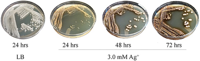
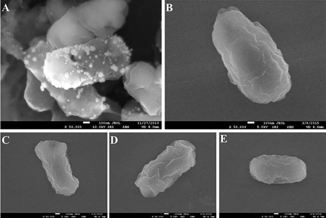
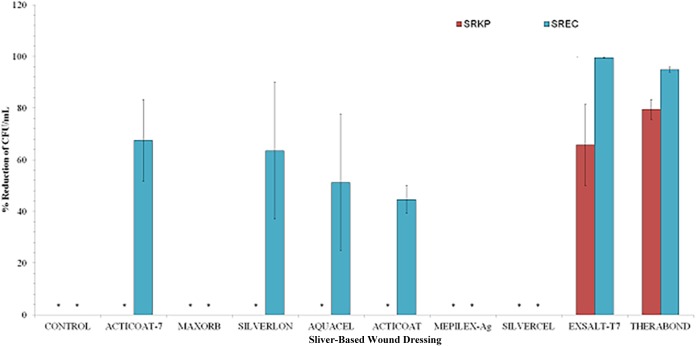
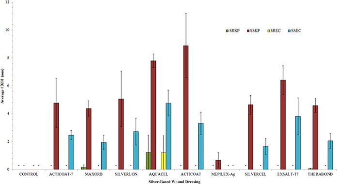
Increased utilization of inorganic silver as an adjunctive to many medical devices has raised concerns of emergent silver resistance in clinical bacteria. Although the molecular basis for silver resistance has been previously characterized, to date, significant phenotypic expression of these genes in clinical settings is yet to be observed. Here, we identified the first strains of clinical bacteria expressing silver resistance at a level that could significantly impact wound care and the use of silver-based dressings. Screening of 859 clinical isolates confirmed 31 harbored at least 1 silver resistance gene. Despite the presence of these genes, MIC testing revealed most of the bacteria displayed little or no increase in resistance to ionic silver (200 to 300 μM Ag(+)). However, 2 isolates (Klebsiella pneumonia and Enterobacter cloacae) were capable of robust growth at exceedingly high silver concentrations, with MIC values reaching 5,500 μM Ag(+). DNA sequencing of these two strains revealed the presence of genes homologous to known genetic determinants of heavy metal resistance. Darkening of the bacteria's pigment was observed after exposure to high silver concentrations. Scanning electron microscopy images showed the presence of silver nanoparticles embedded in the extracellular polymeric substance of both isolates. This finding suggested that the isolates may neutralize ionic silver via reduction to elemental silver. Antimicrobial testing revealed both organisms to be completely resistant to many commercially available silver-impregnated burn and wound dressings. Taken together, these findings provide the first evidence of clinical bacteria capable of expressing silver resistance at levels that could significantly impact wound management.
Copyright © 2015, American Society for Microbiology. All Rights Reserved.
Figures
Similar articles
-
Clinically isolated bacteria resistance to silver-based wound dressings.J Wound Care. 2021 Mar 2;30(3):238-247. doi: 10.12968/jowc.2021.30.3.238. J Wound Care. 2021. PMID: 33729837
-
Prevalence of silver resistance in bacteria isolated from diabetic foot ulcers and efficacy of silver-containing wound dressings.Ostomy Wound Manage. 2008 Mar;54(3):30-40. Ostomy Wound Manage. 2008. PMID: 18382046
-
Prevalence of silver resistance genes in bacteria isolated from human and horse wounds.Vet Microbiol. 2009 Sep 18;138(3-4):325-9. doi: 10.1016/j.vetmic.2009.03.023. Epub 2009 Mar 24. Vet Microbiol. 2009. PMID: 19362435
-
Bacterial resistance to silver in wound care and medical devices.J Wound Care. 2007 Jan;16(1):15-9. doi: 10.12968/jowc.2007.16.1.26983. J Wound Care. 2007. PMID: 17334141 Review.
-
Silver in medicine: the basic science.Burns. 2014 Dec;40 Suppl 1:S9-S18. doi: 10.1016/j.burns.2014.09.010. Burns. 2014. PMID: 25418438 Review.
Cited by
-
Optically Responsive, Smart Anti-Bacterial Coatings via the Photofluidization of Azobenzenes.ACS Appl Mater Interfaces. 2019 Jan 16;11(2):1760-1765. doi: 10.1021/acsami.8b21058. Epub 2019 Jan 4. ACS Appl Mater Interfaces. 2019. PMID: 30605328 Free PMC article.
-
Transcriptional Landscape of a bla KPC-2 Plasmid and Response to Imipenem Exposure in Escherichia coli TOP10.Front Microbiol. 2018 Dec 3;9:2929. doi: 10.3389/fmicb.2018.02929. eCollection 2018. Front Microbiol. 2018. PMID: 30559731 Free PMC article.
-
Multidrug-Resistant CTX-M-(15, 9, 2)- and KPC-2-Producing Enterobacter hormaechei and Enterobacter asburiae Isolates Possessed a Set of Acquired Heavy Metal Tolerance Genes Including a Chromosomal sil Operon (for Acquired Silver Resistance).Front Microbiol. 2018 Mar 23;9:539. doi: 10.3389/fmicb.2018.00539. eCollection 2018. Front Microbiol. 2018. PMID: 29628916 Free PMC article.
-
Nanoparticles as therapeutic options for treating multidrug-resistant bacteria: research progress, challenges, and prospects.World J Microbiol Biotechnol. 2021 May 28;37(6):108. doi: 10.1007/s11274-021-03070-x. World J Microbiol Biotechnol. 2021. PMID: 34046779 Free PMC article. Review.
-
Resistance and Adaptation of Bacteria to Non-Antibiotic Antibacterial Agents: Physical Stressors, Nanoparticles, and Bacteriophages.Antibiotics (Basel). 2021 Apr 13;10(4):435. doi: 10.3390/antibiotics10040435. Antibiotics (Basel). 2021. PMID: 33924618 Free PMC article. Review.
References
-
- Klasen HJ. 2000. Historical review of the use of silver in the treatment of burns. I. Early uses. Burns 26:117–130. http://dx.doi.org/10.1016/S0305-4179(99)00108-4. - DOI - PubMed
MeSH terms
Substances
LinkOut - more resources
Full Text Sources
Medical
Molecular Biology Databases
