Review
. 2016 Apr;6(2):77-95.
doi: 10.1007/s13346-015-0236-0.

Biomaterial-mediated strategies targeting vascularization for bone repair

Affiliations
Review

Biomaterial-mediated strategies targeting vascularization for bone repair

José R García et al. Drug Deliv Transl Res. 2016 Apr.

Abstract

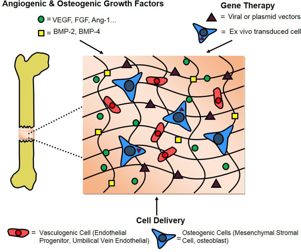
Repair of non-healing bone defects through tissue engineering strategies remains a challenging feat in the clinic due to the aversive microenvironment surrounding the injured tissue. The vascular damage that occurs following a bone injury causes extreme ischemia and a loss of circulating cells that contribute to regeneration. Tissue-engineered constructs aimed at regenerating the injured bone suffer from complications based on the slow progression of endogenous vascular repair and often fail at bridging the bone defect. To that end, various strategies have been explored to increase blood vessel regeneration within defects to facilitate both tissue-engineered and natural repair processes. Developments that induce robust vascularization will need to consolidate various parameters including optimization of embedded therapeutics, scaffold characteristics, and successful integration between the construct and the biological tissue. This review provides an overview of current strategies as well as new developments in engineering biomaterials to induce reparation of a functional vascular supply in the context of bone repair.

Keywords: Bone regeneration; Cell therapy; Growth factor; Tissue engineering; Vascularization.


Conflict of interest statement

Conflicts of Interest

The authors report no conflict of interest

Figures

Figure 1
Figure 1
Current biomaterial-based strategies for inducing vascularization of a critically-sized bone defect
Figure 2
Figure 2
Bioluminescence imaging of Renilla reniformis (RLuc) luciferase under transcriptional control of the cytomegalovirus promoter and PLuc under transcriptional control of the human osteocalcin promoter in transduced human adipose-derived mesenchymal stem cells. Cells were seeded within fibrin matrices and implanted in a 3 mm calvarial defect in SCID mice. A) Representative bioluminescence images showing RLuc (top row) and PLuc (bottom row) intensities. Color bars indicate light intensities from RLuc (black=low; blue=high) and PLuc (blue=low; red=high) B) Quantification of the bioluminescence signal under the constitutive promoter RLuc normalized to day 1. C) Quantification of the bioluminescence signal under the osteocalcin promoter PLuc normalized to RLuc. While osteocalcin signal increases at 2 and 3 weeks, the total amount of cell signal decreases after week one in immunocompromised mice. Adapted from Vila et al [66].
Figure 3
Figure 3
MicroCT angiography of a femoral bone defect treated with rhBMP2 and stabilized with fixation plates that were either locked together to prevent all modes of load transfer (stiff) or unlocked to allow transfer of compressive loads along the bone axis (compliant) at 3 weeks post-surgery. A) Representative 3D reconstructions of vascular networks in total or defect region volume of interest. Scale bar= 1 mm B,C) Vascular volume in total and defect volume of interest, respectively D,E) Vascular connectivity in total and defect volume of interest, respectively. Compliant fixations allowing transfer of mechanical loading exhibited significantly reduced vascular connectivity compared to stiff fixtures. Adapted from Boerckel et al. [146]
Figure 4
Figure 4
Novel biomaterial strategies that hold potential for applications in vascularized bone tissue engineering
Figure 5
Figure 5
Blood vessel labelling and quantification following staining of FTIC-conjugated isolectin b4 from myocardial tissue following a myocardial infarct. A) Representative images (isolectin=green; DAPI=blue; scale bar=30 µm) of rats having either ischemic reperfusion by itself or with delivery of hepatocyte growth factor (HGF) and VEGF from either bolus injection or incorporated within a PEG-maleimide matrix B) Quantification showing increased blood vessel area in PEG/VEGF groups compared to ischemic reperfusion alone and PEG/HGF/VEGF compared to PEG/VEGF and ischemic reperfusion alone. Interestingly, there is no difference between bolus growth factor delivery coupled with ischemic reperfusion and reperfusion alone. Adapted from Salimath et al. [154]
Figure 6
Figure 6
Increased pancreatic beta cell viability and metabolic activity over 3 weeks under low oxygen conditions when incubated with calcium percarbonate containing PDMS disks. A) MTT metabolic activity and B) total DNA quantification. C) Representative images of live/dead staining (green=live; red=dead) of pancreatic beta cells when incubated with or without PDMS-CaO2 materials. (Scale bar= 100 µm). Adapted from Pedraza et al. [175]

Similar articles

Cited by

References

    1. Novosel EC, Kleinhans C, Kluger PJ. Vascularization is the key challenge in tissue engineering. Adv Drug Deliv Rev. 2011;63(4–5):300–311. - PubMed
    1. Logeart-Avramoglou D, Anagnostou F, Bizios R, Petite H. Engineering bone: challenges and obstacles. J Cell Mol Med. 2005;9(1):72–84. - PMC - PubMed
    1. Santos MI, Reis RL. Vascularization in bone tissue engineering: physiology, current strategies, major hurdles and future challenges. Macromol Biosci. 2010;10(1):12–27. - PubMed
    1. Olsen BR, Reginato AM, Wang W. Bone development. Annu Rev Cell Dev Biol. 2000;16:191–220. - PubMed
    1. Akiyama H, Chaboissier MC, Martin JF, Schedl A, de Crombrugghe B. The transcription factor Sox9 has essential roles in successive steps of the chondrocyte differentiation pathway and is required for expression of Sox5 and Sox6. Genes Dev. 2002;16(21):2813–2828. - PMC - PubMed

Publication types

Substances

LinkOut - more resources