. 2015 Jun 9;112(23):7195-200.
doi: 10.1073/pnas.1504177112. Epub 2015 May 26.

Neural Hedgehog signaling maintains stem cell renewal in the sensory touch dome epithelium

Affiliations

Neural Hedgehog signaling maintains stem cell renewal in the sensory touch dome epithelium

Ying Xiao et al. Proc Natl Acad Sci U S A. .

Abstract

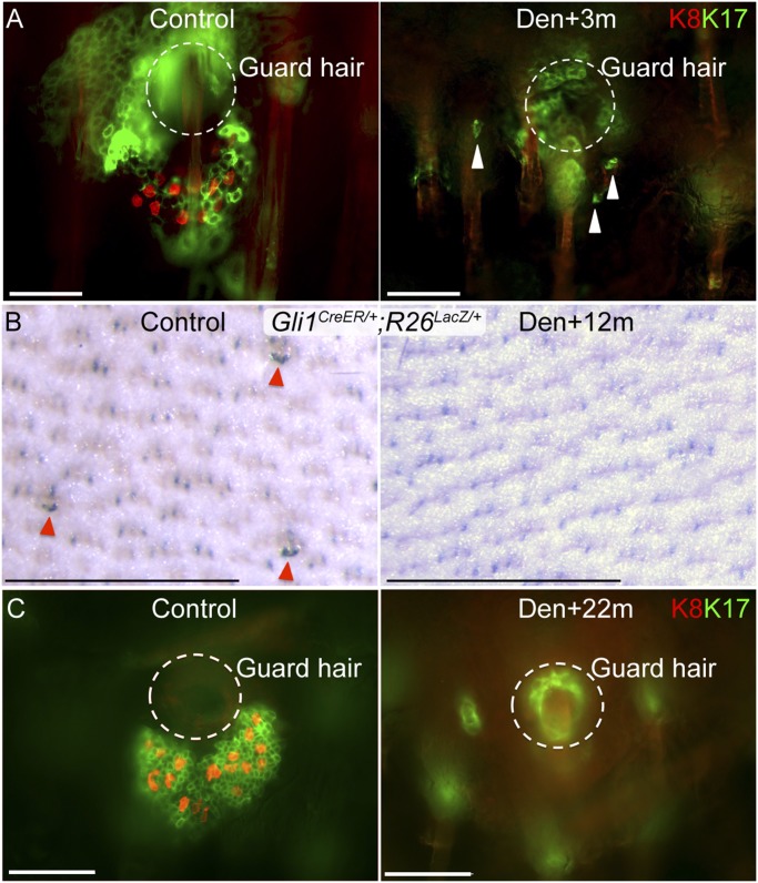
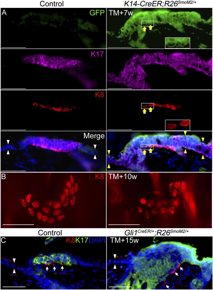
The touch dome is a highly patterned mechanosensory structure in the epidermis composed of specialized keratinocytes in juxtaposition with innervated Merkel cells. The touch dome epithelium is maintained by tissue-specific stem cells, but the signals that regulate the touch dome are not known. We identify touch dome stem cells that are unique among epidermal cells in their activated Hedgehog signaling and ability to maintain the touch dome as a distinct lineage compartment. Skin denervation reveals that renewal of touch dome stem cells requires a perineural microenvironment, and deleting Sonic hedgehog (Shh) in neurons or Smoothened in the epidermis demonstrates that Shh is an essential niche factor that maintains touch dome stem cells. Up-regulation of Hedgehog signaling results in neoplastic expansion of touch dome keratinocytes but no Merkel cell neoplasia. These findings demonstrate that nerve-derived Shh is a critical regulator of lineage-specific stem cells that maintain specialized sensory compartments in the epidermis.

Keywords: Hedgehog; Merkel cell; perineural niche; stem cell; touch dome.


Conflict of interest statement

The authors declare no conflict of interest.

Figures

Fig. 1.
Fig. 1.
Gli1+, Hh-responding stem cells maintain the touch dome in mouse skin. (A and A′) X-Gal whole-mount and section staining in skin from the back of a P59 adult Gli1LacZ/+ mouse. Arrowheads indicate touch domes. (B) β-Galactosidase (β-gal) and K8 staining in skin from the back of an adult Gli1LacZ/+ mouse. (C) X-Gal (false-colored red) with K17 staining in skin from the back of an adult Gli1LacZ/+ mouse. (D) K8 and GFP staining in skin from a Gli1-GIFM (Gli1CreER/+;R26YFP/+) mouse 9 d after tamoxifen induction at P23. Asterisks in B and D indicate nonspecific staining. Yellow arrows in D and E indicate labeled Merkel cells. The red arrows in D indicate unlabeled Merkel cells. (E) K8, K17, and GFP staining in skin from an adult Gli1-GIFM (Gli1CreER/+;R26YFP/+) mouse 3 mo after depilation and tamoxifen induction. (F and F′) X-Gal whole-mount and section staining in skin from the back of a Gli1-GIFM (Gli1CreER/+;R26LacZ/+) mouse 4.5 mo after tamoxifen induction. (G) X-Gal whole-mount staining in skin from adult control and K5-tTA;TRE-cre;Smoflox/flox;Gli1LacZ/+ mice 7 wk after dox withdrawal. (Scale bars, 50 μm for sections; 0.5 mm for whole-mount skin.)
Fig. 2.
Fig. 2.
Shh from sensory nerves is the source of Hh signaling to touch dome stem cells. (A) GFP and K8 staining at the Merkel cell–neurite complex in skin from an adult ShhCreGFP/+;TauLacZ-mGFP/+ mouse. Asterisks indicate nonspecific staining. (B and C) X-Gal whole-mount staining (B) and section staining (C) in denervated (Den) skin from a Gli1LacZ/+ mouse 2 wk after denervation (Right) and in control skin (Left). Dermal pigment and adjacent guard hairs mark touch dome locations. (Insets) Magnified guard hair follicle opening (circles) and adjacent touch dome (arrowheads). (D) X-Gal whole-mount staining in denervated (Right) and control (Left) skin from a Gli1-GIFM (Gli1CreER/+;R26LacZ/+) mouse 4 wk after denervation. (Scale bars, 50 μm for sections; 0.5 mm for whole-mount skin.)
Fig. 3.
Fig. 3.
Surgical denervation causes gradual loss of touch dome. (A) K8 and K17 whole-mount staining in mouse skin 3 mo after denervation (Den). The arrowhead indicates residual K17+ touch dome cells. (B) X-Gal whole-mount staining in skin from the back of a Gli1-GIFM (Gli1CreER/+;R26LacZ/+) mouse 12 mo after surgical denervation. Denervation was performed at 2 wk after tamoxifen induction. Arrowheads indicate touch domes. (C) K8 and K17 whole-mount staining in mouse skin 22 mo after denervation. Circles show the location of the guard follicle based on visualization in a deeper focal plane. (Scale bars, 50 μm for immunofluorescent staining; 0.5 mm for whole-mount X-Gal staining.)
Fig. 4.
Fig. 4.
Nerve-derived Shh signaling is essential for touch dome maintenance. (A) K8 whole-mount staining in P0 WT and Wnt1-Cre;Shhflox/flox mice. (B) K8 and K17 whole-mount staining in P18 WT and Wnt1-Cre;Shhflox/flox mice. (C) The number of K8+ Merkel cells per touch dome (mean ± SEM) in skin from WT and mutant (Mut;, Wnt1-Cre;Shhflox/flox) mice at P18 and P78. (D) Relative expression of Shh mRNA in P16 WT and mutant DRG neurons. (E) The experimental scheme for epidermal deletion of Smo by dox withdrawal. (F) The number of K8+ Merkel cells per touch dome (mean ± SEM) in skin from WT and mutant (Mut; K5-tTA;TRE-cre;Smoflox/flox) mice after Smo deletion at P0 or P21. ***P < 0.0001. (Scale bars, 50 μm.)
Fig. 5.
Fig. 5.
Autonomous activation of Hh signaling in epidermis induces keratinocyte expansion but not Merkel cell hyperplasia. (A) K8, K17, and GFP staining in vehicle-treated (control) and tamoxifen-treated K14-CreER;R26SmoM2/+ mice 7 wk after tamoxifen induction. The white arrowheads indicate normal epidermis. Yellow arrowheads indicate neoplastic epidermis. (Insets) SmoM2-YFP expressing K8+ Merkel cells. Yellow arrows indicate a GFP-labeled Merkel cell. (B) K8 whole-mount staining in control and tamoxifen-treated K14-CreER;R26SmoM2/+ mice 10 wk after tamoxifen induction. (C) K8 and K17 staining in control and Gli1CreER/+;R26SmoM2/+ mice 15 wk after tamoxifen induction. White arrowheads indicate normal epidermis. White arrows indicate K8+ Merkel cells. (Scale bars, 50 μm.)

References

    1. Page ME, Lombard P, Ng F, Göttgens B, Jensen KB. The epidermis comprises autonomous compartments maintained by distinct stem cell populations. Cell Stem Cell. 2013;13(4):471–482. - PMC - PubMed
    1. Liu S, Zhang H, Duan E. Epidermal development in mammals: Key regulators, signals from beneath, and stem cells. Int J Mol Sci. 2013;14(6):10869–10895. - PMC - PubMed
    1. Morgan BA. 2014. The dermal papilla: An instructive niche for epithelial stem and progenitor cells in development and regeneration of the hair follicle. Cold Spring Harb Perspect Med 4(7):a015180.
    1. Gomez C, et al. The interfollicular epidermis of adult mouse tail comprises two distinct cell lineages that are differentially regulated by Wnt, Edaradd, and Lrig1. Stem Cell Reports. 2013;1(1):19–27. - PMC - PubMed
    1. Festa E, et al. Adipocyte lineage cells contribute to the skin stem cell niche to drive hair cycling. Cell. 2011;146(5):761–771. - PMC - PubMed

Publication types

Substances