. 2015 Jun 9;112(23):7219-24.
doi: 10.1073/pnas.1507977112. Epub 2015 May 26.

Human HLA-G+ extravillous trophoblasts: Immune-activating cells that interact with decidual leukocytes

Affiliations

Human HLA-G+ extravillous trophoblasts: Immune-activating cells that interact with decidual leukocytes

Tamara Tilburgs et al. Proc Natl Acad Sci U S A. .

Abstract

Invading human leukocyte antigen-G+ (HLA-G+) extravillous trophoblasts (EVT) are rare cells that are believed to play a key role in the prevention of a maternal immune attack on foreign fetal tissues. Here highly purified HLA-G+ EVT and HLA-G- villous trophoblasts (VT) were isolated. Culture on fibronectin that EVT encounter on invading the uterus increased HLA-G, EGF-Receptor-2, and LIF-Receptor expression on EVT, presumably representing a further differentiation state. Microarray and functional gene set enrichment analysis revealed a striking immune-activating potential for EVT that was absent in VT. Cocultures of HLA-G+ EVT with sample matched decidual natural killer cells (dNK), macrophages, and CD4+ and CD8+ T cells were established. Interaction of EVT with CD4+ T cells resulted in increased numbers of CD4+CD25(HI)FOXP3+CD45RA+ resting regulatory T cells (Treg) and increased the expression level of the Treg-specific transcription factor FOXP3 in these cells. However, EVT did not enhance cytokine secretion in dNK, whereas stimulation of dNK with mitogens or classical natural killer targets confirmed the distinct cytokine secretion profiles of dNK and peripheral blood NK cells (pNK). EVT are specialized cells involved in maternal-fetal tolerance, the properties of which are not imitated by HLA-G-expressing surrogate cell lines.

Keywords: FOXP3; NK cell; Treg; human; pregnancy.


Conflict of interest statement

Conflict of interest statement: J.L.S. is a consultant for King Abdulaziz University, Jeddah, Saudi Arabia.

Figures
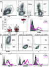
Fig. 1.
Fig. 1.
Trophoblast preparations. (A) Representative FACS plots of trophoblast preparations. A live gate (R1) was set in the Forward-Side Scatter plot and CD45+ leukocytes were excluded with gate R2. Subsequently, EGFR1–FITC and HLA-G–PE were plotted, and separation of EVT (CD45-EGFR1dimHLA-G+) and VT (CD45-HLA-G-EGFR1+) is depicted. (B) Percentage of CD45+ leukocytes (within live gate), CD45-EGFR1dimHLA-G+ EVT, and CD45-HLA-G-EGFR1+ VT (both within live gate and CD45− fraction) of the trophoblast preparations. (C) HLA-G expression on freshly isolated trophoblast preparations and preparations cultured for 2 d on fibronectin. (D) Representative FACS plots of HLA-G and EGFR1 expression on FACS-sorted CD45-HLA-G+ EVT, CD45-HLA-G-EGFR1+ VT, and the remaining CD45-HLA-G-EGFR1− cells that were cultured on fibronectin for 2 d. (E) EGFR1, EGFR2, and LIFR expression on freshly isolated EVT and EVT cultured for 2 d on fibronectin.
Fig. 2.
Fig. 2.
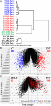
Expression profiling of EVT and VT. (A) Unsupervised hierarchical cluster analysis results in a dendrogram where each distinct cell type (VT, EVT, JEG-3, and DSC) forms a separate cluster. Volcano plots were generated based on the mean expression value of an individual probe’s fold change and the P value associated with reproducibility of these changes between (B) VT and EVT and (C) EVT and JEG-3. The unique gene signatures based on a more than fourfold difference for VT (340 probes, blue dots) and EVT (328 probes, red dots) are highlighted.
Fig. 3.
Fig. 3.
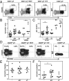
Coculture with EVT increases the percentage of CD4+FOXP3+ Treg. (A) Representative FACS plots of CD25 and FOXP3 expression of CD4+ pT in the absence of stimulation or after coculture with VT- or EVT-enriched preparations. (B) The percentage of FOXP3+ cells within total CD4+ pT and dT and (C) MFI of FOXP3 expression within CD4+CD25HIFOXP3+ pT and dT cocultured with or without VT- or EVT-enriched preparations. (D) Representative FACS plots of CD45RA and FOXP3 within CD4+CD25hi pT cocultured with or without VT- or EVT-enriched preparations. (E) The percentage of FOXP3+CD45RA− and (F) FOXP3+CD45RA+ cells within each group.
Fig. 4.
Fig. 4.
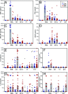
Cytokine secretion profiles of pNK and dNK during coculture with VT and EVT. pNK (blue) and dNK (red) were incubated alone or with PMA, 221, 221/HLA-G, and VT- or EVT-enriched preparations for 18 h. Cell culture supernatants were analyzed for (A) GM-CSF, (B) IFNγ, (C) TNFα, (D) VEGF, (E) IL-6, (F) IL-17A, (G) IL-8, and (H) galectin-1. VT- or EVT-enriched preparations cultured alone secreted IL-6 and IL-8 (E and G, depicted by gray dots), but did not secrete any of the other cytokines.

References

    1. Moffett A, Loke C. Immunology of placentation in eutherian mammals. Nat Rev Immunol. 2006;6(8):584–594. - PubMed
    1. Jokhi PP, King A, Loke YW. Reciprocal expression of epidermal growth factor receptor (EGF-R) and c-erbB2 by non-invasive and invasive human trophoblast populations. Cytokine. 1994;6(4):433–442. - PubMed
    1. Kimber SJ. Leukaemia inhibitory factor in implantation and uterine biology. Reproduction. 2005;130(2):131–145. - PubMed
    1. Das P, et al. Effects of fgf2 and oxygen in the bmp4-driven differentiation of trophoblast from human embryonic stem cells. Stem Cell Res (Amst) 2007;1(1):61–74. - PMC - PubMed
    1. Roberts RM, Fisher SJ. Trophoblast stem cells. Biol Reprod. 2011;84(3):412–421. - PMC - PubMed

Publication types