. 2015 Jun 9;112(23):7189-94.
doi: 10.1073/pnas.1504822112. Epub 2015 May 26.

The disordered P granule protein LAF-1 drives phase separation into droplets with tunable viscosity and dynamics

Affiliations

The disordered P granule protein LAF-1 drives phase separation into droplets with tunable viscosity and dynamics

Shana Elbaum-Garfinkle et al. Proc Natl Acad Sci U S A. .

Abstract

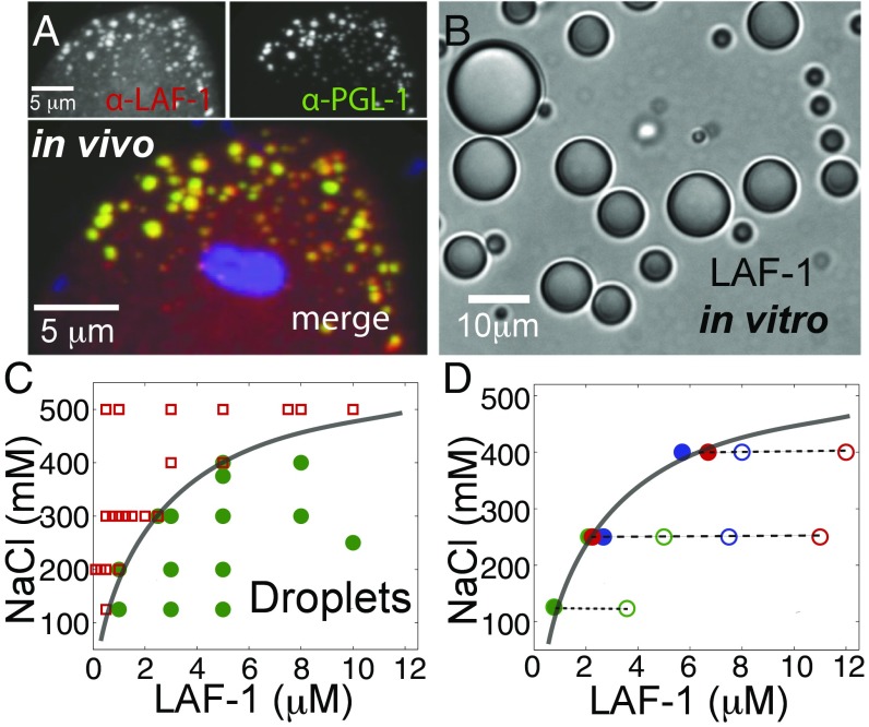
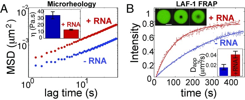
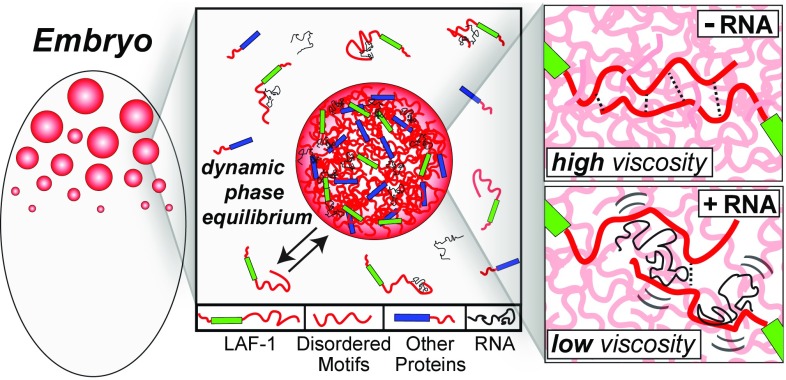
P granules and other RNA/protein bodies are membrane-less organelles that may assemble by intracellular phase separation, similar to the condensation of water vapor into droplets. However, the molecular driving forces and the nature of the condensed phases remain poorly understood. Here, we show that the Caenorhabditis elegans protein LAF-1, a DDX3 RNA helicase found in P granules, phase separates into P granule-like droplets in vitro. We adapt a microrheology technique to precisely measure the viscoelasticity of micrometer-sized LAF-1 droplets, revealing purely viscous properties highly tunable by salt and RNA concentration. RNA decreases viscosity and increases molecular dynamics within the droplet. Single molecule FRET assays suggest that this RNA fluidization results from highly dynamic RNA-protein interactions that emerge close to the droplet phase boundary. We demonstrate than an N-terminal, arginine/glycine rich, intrinsically disordered protein (IDP) domain of LAF-1 is necessary and sufficient for both phase separation and RNA-protein interactions. In vivo, RNAi knockdown of LAF-1 results in the dissolution of P granules in the early embryo, with an apparent submicromolar phase boundary comparable to that measured in vitro. Together, these findings demonstrate that LAF-1 is important for promoting P granule assembly and provide insight into the mechanism by which IDP-driven molecular interactions give rise to liquid phase organelles with tunable properties.

Keywords: RNA granules; intracellular phase transition; intrinsically disordered proteins; liquid droplets.


Conflict of interest statement

The authors declare no conflict of interest.

Figures

Fig. 1.
Fig. 1.
LAF-1 colocalizes to P granules in vivo and phase separates into droplets in vitro. (A) Confocal images of two-cell embryo posterior immunostained for LAF-1 (Upper Left) and PGL-1 (Upper Right). In the dividing P1 cell, LAF-1 localizes to PGL-1–marked P granules; DAPI-stained nucleus is included in the merged image. (B) DIC image of phase separated LAF-1 droplets. (C) Protein/NaCl concentrations scoring positive (green circles) or negative (red squares) for optically resolvable droplets are plotted, resulting in a phase boundary (line drawn to guide the eye). (D) The protein concentration in the dilute phase (●) is plotted for varying total protein concentrations (○) at three different salt concentrations. For all conditions, the concentration of the dilute phase falls directly onto the LAF-1 phase boundary from C (solid line).
Fig. 2.
Fig. 2.
LAF-1 droplets are homogeneous fluids with salt-dependent viscosity. (A) Confocal image sequence showing LAF-1 droplet fusion. (B) Fusion events are well fit by an exponential decay, which is used to determine fusion timescale τ. (C) Decay time vs. length scale for LAF-1 droplets prepared in 125 mM NaCl. The linear slope represents the inverse capillary velocity, η/γ0.12 s/μm. (D) Confocal image of red fluorescent beads embedded inside a large LAF-1 droplet. (E) Probability distribution of bead displacement for three different lag times. Distributions are well fit to a Gaussian (solid lines) indicating a homogenous environment. (F) Mean squared displacement vs. lag time. MSD data for individual beads from a single droplet are plotted. Black solid line, slope of 1; black dash, the noisefloor is 2 × 10⁻⁴ μm. (Inset) Representative 2D particle track. (G) Increasing concentrations of NaCl result in increased MSD of particles and decreased viscosity (Inset).
Fig. 3.
Fig. 3.
RNA fluidizes LAF-1 droplets. (A) RNA addition (5 μM pU50) increases MSD of particles in LAF-1 droplets and decreases viscosity: (Inset) [NaCl] = 125 mM. (B) The timescale of LAF-1 FRAP recovery within droplets decreases on addition of 5 μM pU50 RNA (red). (Upper Inset) Representative droplets in which 1% of LAF-1 is labeled with DyLight 488: prebleach (Left), postbleach (Center), and t 300 s (Right). (Lower Inset) Calculated apparent diffusion coefficients.
Fig. 4.
Fig. 4.
LAF-1–induced RNA dynamics activated near phase boundary. (A) LAF-1–RNA interactions are measured across the phase boundary by increasing the protein concentration (green arrow) and decreasing the NaCl concentration (red arrow). Measurements were performed at the indicated LAF-1 and NaCl concentrations (colored circles). (B) As the concentration of LAF-1 approaches the phase boundary, the peak at FRET values of 0.8 decreases, shifts, and broadens (Left). Single molecule FRET efficiency traces reveal dynamic pattern of LAF-1–RNA interactions emerging at increasing concentration (Right). [NaCl] held constant at 125 mM. (C) As the NaCl concentration approaches and crosses the phase boundary, the FRET peak similarly shifts and broadens (Left), concomitant with increased FRET dynamics (Right).
Fig. 5.
Fig. 5.
Disordered N-terminal RGG domain drives phase separation and RNA/protein dynamics. (A) The PONDR algorithm predicts a high degree of disorder for both N and C termini of LAF-1. (B) Circular dichroism spectra indicate that the N-terminal RGG domain resembles a random coil, whereas FL, ΔC, and ΔRGG constructs contain secondary structure dominated by alpha helical conformation. (C) Schematic of LAF-1 deletion constructs. (Right) Droplets formed in 125 mM NaCl with DyLight-488–labeled protein for the indicated LAF-1 construct. (D) Single molecule FRET traces were recorded for 2 μM protein and 125 mM NaCl. The RGG domain is necessary and sufficient for dynamic RNA–protein interactions.
Fig. 6.
Fig. 6.
LAF-1 depletion disrupts P granule organization. (A) The percentage of hatched embryos laid by singled hermaphrodites (n = 42) is strongly decreased in laf-1(RNAi) mothers. (B) Estimation of LAF-1 concentration in the germplasm as a function of laf-1(RNAi) exposure. (C–E) Epifluorescent images of four-cell embryos immunostained for LAF-1 (C), PGL-1 (D), and PIE-1 (E) at 30 h of RNAi feeding. (Scale bars, 10 μm.)
Fig. 7.
Fig. 7.
Schematic illustrating the role of LAF-1 IDP motifs and RNA in droplet assembly and properties. IDP motifs (red) in LAF-1 (green) and likely other P granule proteins (blue) are important for driving phase separation into dynamic but coherent liquid droplets. These droplets are a liquid phase but with relatively high viscosity. RNA gives rise to dynamic interactions with LAF-1 (and likely other) IDP domains, modulating IDP–IDP interactions and leading to decreased droplet viscosity and increased molecular dynamics within the droplet.

References

    1. Anderson P, Kedersha N. RNA granules. J Cell Biol. 2006;172(6):803–808. - PMC - PubMed
    1. Brangwynne CP, et al. Germline P granules are liquid droplets that localize by controlled dissolution/condensation. Science. 2009;324(5935):1729–1732. - PubMed
    1. Brangwynne CP, Mitchison TJ, Hyman AA. Active liquid-like behavior of nucleoli determines their size and shape in Xenopus laevis oocytes. Proc Natl Acad Sci USA. 2011;108(11):4334–4339. - PMC - PubMed
    1. Wippich F, et al. Dual specificity kinase DYRK3 couples stress granule condensation/dissolution to mTORC1 signaling. Cell. 2013;152(4):791–805. - PubMed
    1. Feric M, Brangwynne CP. A nuclear F-actin scaffold stabilizes RNP droplets against gravity in large cells. Nat Cell Biol. 2013;15(10):1253–1259. - PMC - PubMed

Publication types

MeSH terms