

Effect of distance and duration of illumination on retinal ganglion cells exposed to varying concentrations of brilliant blue green
- PMID: 26015816
- PMCID: PMC4432893
- DOI: 10.14740/jocmr2085e
Effect of distance and duration of illumination on retinal ganglion cells exposed to varying concentrations of brilliant blue green
Abstract
Background: The objective of the study was to determine the safety parameters of using brilliant blue green (BBG) for chromovitrectomy by assessing the cytotoxicity of BBG on cultured retinal ganglion cells (RGCs) exposed to illumination.
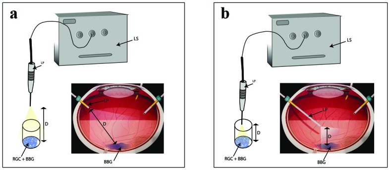
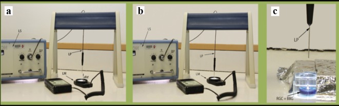
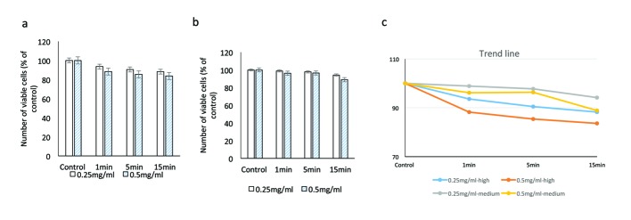
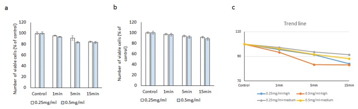
Methods: RGCs were exposed to two concentrations of BBG (0.25 and 0.5 mg/mL) under metal halide illumination at varying distances (1 and 2.5 cm), intensities (990 and 2,000 Fc), and durations (1, 5 and 15 minutes). Cell viability was assessed using the WST-1 and CellTiter 96(®) AQueous One solution cell proliferation assays.
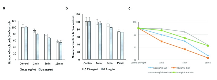
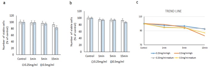
Results: Using the WST-1 assay, with high-intensity illumination, viability of RGCs ranged from 97.5±16.4% of controls with minimum BBG and light exposure (0.25 mg/mL BBG and illuminated for 1 minute at 2.5 cm distance) to 53.1±11.3% of controls with maximum BBG and light exposure (0.50 mg/mL and illuminated for 15 minutes at 1 cm distance; P < 0.01). With medium-intensity illumination, RGCs showed better viability, ranging from 95.1±7.2% of controls with minimum BBG and light exposure to 72.3±12.8% of controls with maximum BBG and light exposure. CellTiter 96(®) AQueous One assay showed similar results.
Conclusion: RGCs seem to safely tolerate up to 5 minutes of exposure to 0.5 mg/mL BBG under diffuse medium-intensity illumination (990 Fc).
Keywords: Brilliant blue green; Chromovitrectomy; Cytotoxicity; Endoillumination; Retinal ganglion cells.
Figures
Similar articles
-
Assessment of the effect of distance and duration of illumination on retinal pigment epithelial cells exposed to varying doses of Brilliant Blue Green.J Ocul Pharmacol Ther. 2014 Oct;30(8):625-33. doi: 10.1089/jop.2013.0225. Epub 2014 Jul 7. J Ocul Pharmacol Ther. 2014. PMID: 24999985
-
Osmolarity and spectrophotometric property of brilliant blue green define the degree of toxicity on retinal pigment epithelial cells exposed to surgical endoilluminator.Clin Ophthalmol. 2016 Aug 16;10:1543-51. doi: 10.2147/OPTH.S110930. eCollection 2016. Clin Ophthalmol. 2016. PMID: 27574394 Free PMC article.
-
Comparative in vitro safety analysis of dyes for chromovitrectomy: indocyanine green, brilliant blue green, bromophenol blue, and infracyanine green.Retina. 2011 Jun;31(6):1128-36. doi: 10.1097/IAE.0b013e3181fe543a. Retina. 2011. PMID: 21394068
-
Brilliant blue in vitreoretinal surgery.Dev Ophthalmol. 2008;42:115-125. doi: 10.1159/000138989. Dev Ophthalmol. 2008. PMID: 18535385 Review.
-
[Comprehensive strategy for retinal neuroprotection. Challenging the clinical application].Nippon Ganka Gakkai Zasshi. 2012 Mar;116(3):165-98; discussion 199. Nippon Ganka Gakkai Zasshi. 2012. PMID: 22568101 Review. Japanese.
Cited by
-
Macular peeling-induced retinal damage: clinical and histopathological evaluation after using different dyes.Graefes Arch Clin Exp Ophthalmol. 2018 Sep;256(9):1573-1580. doi: 10.1007/s00417-018-4029-2. Epub 2018 Jun 8. Graefes Arch Clin Exp Ophthalmol. 2018. PMID: 29948176 Clinical Trial.
References
-
- Brooks HL, Jr. Macular hole surgery with and without internal limiting membrane peeling. Ophthalmology. 2000;107(10):1939–1948. discussion 1948-1939. - PubMed
-
- Haritoglou C, Gandorfer A, Gass CA, Schaumberger M, Ulbig MW, Kampik A. Indocyanine green-assisted peeling of the internal limiting membrane in macular hole surgery affects visual outcome: a clinicopathologic correlation. Am J Ophthalmol. 2002;134(6):836–841. doi: 10.1016/S0002-9394(02)01816-0. - DOI - PubMed
LinkOut - more resources
Full Text Sources
Other Literature Sources