. 2015 Jun 9;112(23):E3058-66.
doi: 10.1073/pnas.1503832112. Epub 2015 May 27.

Structure of a bacterial toxin-activating acyltransferase

Affiliations

Structure of a bacterial toxin-activating acyltransferase

Nicholas P Greene et al. Proc Natl Acad Sci U S A. .

Abstract

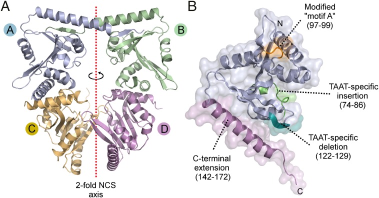
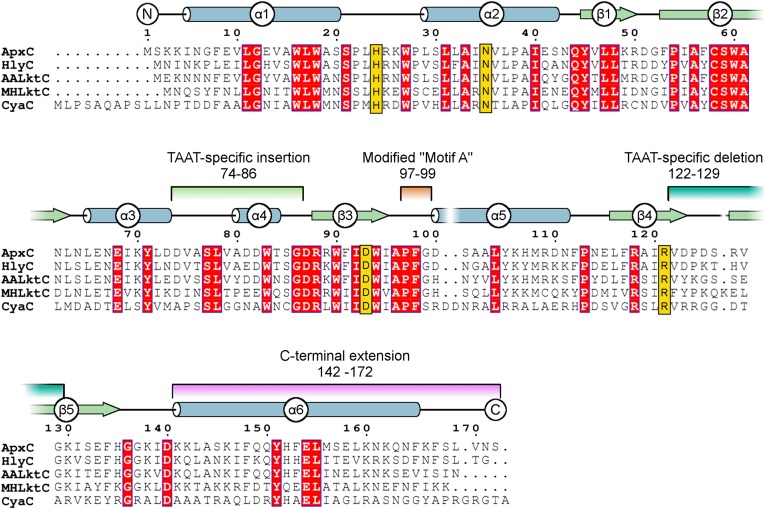
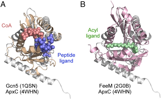
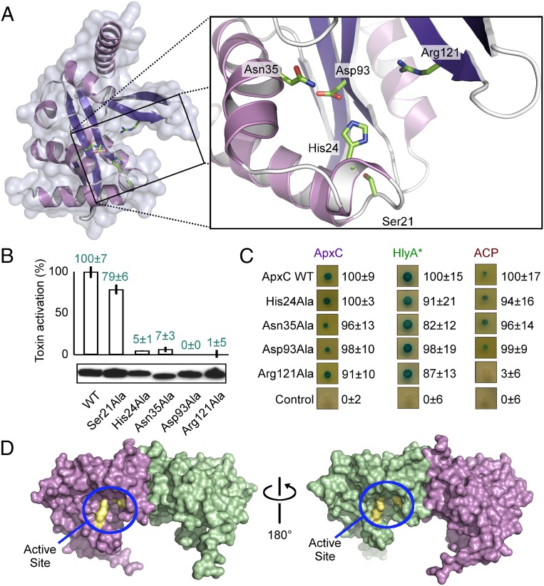
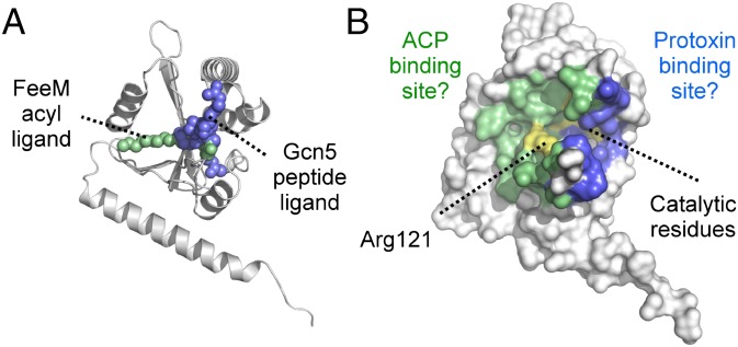
Secreted pore-forming toxins of pathogenic Gram-negative bacteria such as Escherichia coli hemolysin (HlyA) insert into host-cell membranes to subvert signal transduction and induce apoptosis and cell lysis. Unusually, these toxins are synthesized in an inactive form that requires posttranslational activation in the bacterial cytosol. We have previously shown that the activation mechanism is an acylation event directed by a specialized acyl-transferase that uses acyl carrier protein (ACP) to covalently link fatty acids, via an amide bond, to specific internal lysine residues of the protoxin. We now reveal the 2.15-Å resolution X-ray structure of the 172-aa ApxC, a toxin-activating acyl-transferase (TAAT) from pathogenic Actinobacillus pleuropneumoniae. This determination shows that bacterial TAATs are a structurally homologous family that, despite indiscernible sequence similarity, form a distinct branch of the Gcn5-like N-acetyl transferase (GNAT) superfamily of enzymes that typically use acyl-CoA to modify diverse bacterial, archaeal, and eukaryotic substrates. A combination of structural analysis, small angle X-ray scattering, mutagenesis, and cross-linking defined the solution state of TAATs, with intermonomer interactions mediated by an N-terminal α-helix. Superposition of ApxC with substrate-bound GNATs, and assay of toxin activation and binding of acyl-ACP and protoxin peptide substrates by mutated ApxC variants, indicates the enzyme active site to be a deep surface groove.

Keywords: X-ray crystallography; acyl carrier protein; acyltransferase; hemolysin; posttranslational modification.


Conflict of interest statement

The authors declare no conflict of interest.

Figures

Fig. 1.
Fig. 1.
Crystal structure of the toxin-activating acyl transferase ApxC. (A) The asymmetric unit contains four monomers of ApxC. An imperfect twofold noncrystallographic symmetry axis (dotted red line) relates two pairs of monomers. (B) Structure of a single monomer indicating structural features that discern TAATs from other members of the GNAT superfamily.
Fig. 2.
Fig. 2.
Multiple sequence alignment for key members of the TAAT family. Secondary structure of ApxC (consensus of the four monomers in the crystal structure) is shown immediately above the alignment of TAAT sequences ApxC (A. pleuropneumoniae), HlyC (Uropathogenic E. coli), AaLktC (A. actinomycetemcomitans), MhLktC (M. hemolytica), and CyaC (Bordetella pertussis). The α-helices indicated are shown as tubes (blue); β-strands by arrows (green). Conserved residues are highlighted red, and important active site residues identified later (His24, Asn35, Asp93, and Arg121) are shown in gold. Features that differentiate TAATs from other GNATs are highlighted by using the same color scheme as in Fig. 1B.
Fig. 3.
Fig. 3.
X-ray analysis of potential ApxC dimers in crystallo and in solution. (A) The four dimeric arrangements of ApxC extracted from the crystal structure (I–IV) indicating buried surface area and predicted free energy of dissociation (∆G, positive values indicate favorable association) obtained from PISA (34). (B) SAXS analysis of ApxC in solution. (i) The measured SAXS scattering intensity profile is shown in blue (56 μM ApxC) and the predicted scattering profile of the solution model in black, whereas the solution structure of ApxC, a filtered model derived from the average of 20 models, is shown in Inset. (ii) Distance distribution analysis of each of the candidate ApxC dimers verses the SAXS data. The quality of the fit can be seen by comparing predicted P(r) profile in black with the measured profile in blue, quantified by the χ2 value shown in Inset.
Fig. 4.
Fig. 4.
Experimental determination of the ApxC solution dimer. (A) Locations of engineered cysteine residues (marked with spheres Gly12, blue; Lys160, orange) with respect to two dimeric arrangements (dimer III, Left; dimer I, Right). (B) Cysteine cross-linking of purified wild-type (WT) ApxC or cysteine variant. Protein (50 µg) was treated with or without 100 µM CuCl2 (X-linker) for 30 min at 25 °C to catalyze cross-linking of proximal cysteines before blocking free cysteines with 5 mM N-ethylmaleimide (NEM). Preblock reactions were treated with NEM and EDTA before addition of cross-linker. (C) Locations of Gly12, Ala15, and Ala19 within the N-terminal α-helix at the dimer III interface. (D) Bacterial two-hybrid assay of homodimerization of WT ApxC or indicated variants. Interaction was assessed qualitatively by blue coloration and quantitatively by β-galactosidase activity (Right), expressed as a percentage of the wild type ± SD. (E) Location of an engineered C-terminal deletion (Δ162–172) in context of dimer I. (F) Bacterial two-hybrid assay assessing homodimerization of ApxC (Δ162–172), details as for D.
Fig. 5.
Fig. 5.
Superposition of the TAAT ApxC with Gcn5 and FeeM. (A) Superposition of ApxC (white) with Gcn5 (wheat). The two Gcn5 substrates are CoA (red) and histone H3 peptide (blue). (B) Superposition of ApxC (white) with FeeM (pink). The FeeM N-lauroylayrosine ligand is shown in green.
Fig. 6.
Fig. 6.
Definition of the TAAT active site. (A) Location of the postulated substrate-binding groove and close-up of the active site (boxed). Side chains of key active site residues are shown in stick representation. (B) TAAT activity of active site variants assayed by in vivo toxin activation (erythrocyte lysis, percentage of wild-type activity ± SD with immunoblot showing expression underneath. (C) Bacterial two-hybrid experiments testing ApxC dimerization (Left), interaction with TAAT-binding protoxin peptide (residues 679–736 - HlyA*; Center), and ACP binding (Right) for wild-type (WT) ApxC and mutant variants. Interaction was assessed qualitatively by blue coloration and numerically by β-galactosidase activity (Right), expressed as a percentage of the wild type (Top) ± SD after subtraction of the negative control. (D) Solid surface representations of ApxC showing the location of the active site of each monomer in context of the soluble dimer (blue lasso), with important active site residues in yellow.
Fig. 7.
Fig. 7.
A model for ternary complex TAAT function. (A) Location of the acyltyrosine ligand (green) from the FeeM:acyltyrosine costructure and histone H3 peptide (blue) from the Gcn5:CoA:H3 peptide ternary complex after superposition with ApxC (white). (B) Proposed locations of the acylACP and protoxin binding sites on opposite faces of the TAAT enzyme (ACP-binding site green; protoxin binding site blue). The substrates are predicted to meet at the crux of the split β-sheet where the catalytic residues (yellow) are located. Arg121 (essential for ACP binding) is annotated.

References

    1. Ulett GC, et al. Uropathogenic Escherichia coli virulence and innate immune responses during urinary tract infection. Curr Opin Microbiol. 2013;16(1):100–107. - PubMed
    1. Gur C, et al. Natural killer cell-mediated host defense against uropathogenic E. coli is counteracted by bacterial hemolysinA-dependent killing of NK cells. Cell Host Microbe. 2013;14(6):664–674. - PMC - PubMed
    1. Uhlén P, et al. Alpha-haemolysin of uropathogenic E. coli induces Ca2+ oscillations in renal epithelial cells. Nature. 2000;405(6787):694–697. - PubMed
    1. Stanley P, Koronakis V, Hughes C. Acylation of Escherichia coli hemolysin: A unique protein lipidation mechanism underlying toxin function. Microbiol Mol Biol Rev. 1998;62(2):309–333. - PMC - PubMed
    1. Wiles TJ, Dhakal BK, Eto DS, Mulvey MA. Inactivation of host Akt/protein kinase B signaling by bacterial pore-forming toxins. Mol Biol Cell. 2008;19(4):1427–1438. - PMC - PubMed

Publication types

MeSH terms

Substances

Associated data

LinkOut - more resources