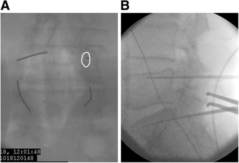
Lumbosacral pedicle screw placement using a fluoroscopic pedicle axis view and a cannulated tapping device
- PMID: 26016564
- PMCID: PMC4450829
- DOI: 10.1186/s13018-015-0225-5
Lumbosacral pedicle screw placement using a fluoroscopic pedicle axis view and a cannulated tapping device
Abstract
Background: Pedicle screw insertions are commonly used for posterior fixation to treat various spine disorders. However, the misplacement of pedicle screws can lead to disastrous complications. Inaccurate pedicle screw placement is relatively common even when placement is performed under fluoroscopic control. In order to improve the accuracy of the screw placement, we applied a technique using guide wires and a cannulated tapping device with the assistance of a fluoroscopic pedicle axis view.
Methods: From 2006 to 2011, 854 pedicle screws were placed in 176 patients in lumbosacral spinal fusion surgeries. The accuracy of screw placement was evaluated using postoperative reconstructed computed tomography images. Screw misplacement was classified as minor (cortical perforation <3 mm), moderate (cortical perforation 3-6 mm), or severe (cortical perforation >6 mm). Using logistic regression analysis, we also investigated the potential risk factors associated with screw misplacement.
Results: Pedicle screw misplacement was observed in 37 screws (4.3%) in 34 patients. In the sub-classification analysis, 28 screws (3.3%) were determined to be minor perforations, 7 screws (0.8%) were considered to be moderate perforations, and 2 screws (0.2%) was judged to be a severe perforation (cortical perforation >6 mm). None of the 28 screws that were considered to be minor perforations were associated with any significant symptoms in the patients. However, 2 of the 9 screws that were determined to be moderate or severe perforations caused neurological symptoms (1 of which required revision). No significant differences were observed in the incidence of screw misplacement among the vertebral levels. Significant risk factors for screw misplacement were obesity and degenerative scoliosis. The odds ratios of these significant risk factors were 3.593 (95% confidence interval (CI), 1.061-12.175) for obesity and 8.893 for degenerative scoliosis (95% CI, 1.200-76.220).
Conclusions: A modified fluoroscopic technique using a pedicle axis view and a cannulated tapping instrument can achieve safe and accurate pedicle screw placement. In addition, obesity and degenerative scoliosis were identified as significant risk factors for screw misplacement.
Figures


Similar articles
-
Accuracy of pedicle screw placement in the thoracic and lumbosacral spine using a conventional intraoperative fluoroscopy-guided technique: a national neurosurgical education and training center analysis of 1236 consecutive screws.World Neurosurg. 2014 Nov;82(5):866-71.e1-2. doi: 10.1016/j.wneu.2014.06.023. Epub 2014 Jun 17. World Neurosurg. 2014. PMID: 24954252
-
Robotic versus fluoroscopy-guided pedicle screw insertion for metastatic spinal disease: a matched-cohort comparison.Neurosurg Focus. 2017 May;42(5):E13. doi: 10.3171/2017.3.FOCUS1710. Neurosurg Focus. 2017. PMID: 28463620
-
Accuracy of fluoroscopically-assisted pedicle screw placement: analysis of 1,218 screws in 198 patients.Spine J. 2014 Aug 1;14(8):1702-8. doi: 10.1016/j.spinee.2014.03.044. Epub 2014 Apr 4. Spine J. 2014. PMID: 24704680
-
Accuracy and Safety of Pedicle Screw Placement in Adolescent Idiopathic Scoliosis Patients: A Review of 2020 Screws Using Computed Tomography Assessment.Spine (Phila Pa 1976). 2017 Mar;42(5):326-335. doi: 10.1097/BRS.0000000000001738. Spine (Phila Pa 1976). 2017. PMID: 27310021 Review.
-
Accuracy of Current Techniques for Placement of Pedicle Screws in the Spine: A Comprehensive Systematic Review and Meta-Analysis of 51,161 Screws.World Neurosurg. 2019 Jun;126:664-678.e3. doi: 10.1016/j.wneu.2019.02.217. Epub 2019 Mar 15. World Neurosurg. 2019. PMID: 30880208
Cited by
-
The sacral screw placement depending on morphological and anatomical peculiarities.Surg Radiol Anat. 2020 Mar;42(3):299-305. doi: 10.1007/s00276-019-02373-x. Epub 2019 Nov 23. Surg Radiol Anat. 2020. PMID: 31760529
-
Accuracy of pedicle screw insertion for unilateral open transforaminal lumbar interbody fusion: a side-by-side comparison of percutaneous and conventional open techniques in the same patients.BMC Musculoskelet Disord. 2020 Mar 14;21(1):168. doi: 10.1186/s12891-020-3180-1. BMC Musculoskelet Disord. 2020. PMID: 32171291 Free PMC article.
-
Automatic pedicle screw planning using atlas-based registration of anatomy and reference trajectories.Phys Med Biol. 2019 Aug 21;64(16):165020. doi: 10.1088/1361-6560/ab2d66. Phys Med Biol. 2019. PMID: 31247607 Free PMC article.
-
Proximal facet joint violation and breaches after percutaneous insertion of 311 lumbar pedicle screws using the pedicle axis fluoroscopic view.Brain Spine. 2025 May 5;5:104274. doi: 10.1016/j.bas.2025.104274. eCollection 2025. Brain Spine. 2025. PMID: 40476153 Free PMC article.
-
Investigation of Radiation Exposure of Medical Staff During Lateral Fluoroscopy for Posterior Spinal Fusion Surgery.J Clin Med. 2024 Oct 27;13(21):6442. doi: 10.3390/jcm13216442. J Clin Med. 2024. PMID: 39518581 Free PMC article.
References
-
- Gelalis ID, Paschos NK, Pakos EE, Politis AN, Arnaoutoglou CM, Karageorgos AC, et al. Accuracy of pedicle screw placement: a systematic review of prospective in vivo studies comparing free hand, fluoroscopy guidance and navigation techniques. Eur Spine J. 2012;21:247–55. doi: 10.1007/s00586-011-2011-3. - DOI - PMC - PubMed
Publication types
MeSH terms
LinkOut - more resources
Full Text Sources
Other Literature Sources

