

Efficient Mapping of Sulfated Glycotopes by Negative Ion Mode nanoLC-MS/MS-Based Sulfoglycomic Analysis of Permethylated Glycans
- PMID: 26016788
- PMCID: PMC4843773
- DOI: 10.1021/acs.analchem.5b01409
Efficient Mapping of Sulfated Glycotopes by Negative Ion Mode nanoLC-MS/MS-Based Sulfoglycomic Analysis of Permethylated Glycans
Abstract
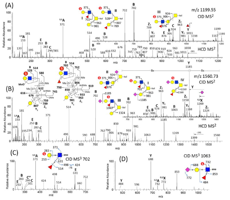
We have previously developed the enabling techniques for sulfoglycomics based on mass spectrometry (MS) analysis of permethylated glycans, which preserves the attractive features of more reliable MS/MS sequencing compared with that performed on native glycans, while providing an easy way to separate and hence enrich the sulfated glycans. Unlike LC-MS/MS analysis of native glycans in negative ion mode that has been more widely in use, the characteristics and potential benefits of similar applications based on permethylated sulfated glycans have not been fully investigated. We report here the important features of reverse phase-based nanoLC-MS/MS analysis of permethylated sulfated glycans in negative ion mode and demonstrate that complementary sets of diagnostic fragment ions afforded can allow rapid identification of various fucosylated, sialylated, sulfated glycotopes and definitive determination of the location of sulfate in a way difficult to achieve by other means. A parallel acquisition of both higher collision energy and trap-based MS(2) coupled with a product dependent MS(3) is conceivably the most productive sulfoglycomic workflow currently possible and the manually curated fragmentation characteristics presented here will allow future developments in automating data analysis.
Conflict of interest statement
Figures


Similar articles
-
Discovery Sulfoglycomics and Identification of the Characteristic Fragment Ions for High-Sensitivity Precise Mapping of Adult Zebrafish Brain-Specific Glycotopes.Front Mol Biosci. 2021 Dec 20;8:771447. doi: 10.3389/fmolb.2021.771447. eCollection 2021. Front Mol Biosci. 2021. PMID: 34988116 Free PMC article.
-
Negative Ion Mode nanoLC-ESI-MS/MS Analyses of Permethylated Sulfated Glycans.Bio Protoc. 2020 May 20;10(10):e3618. doi: 10.21769/BioProtoc.3618. eCollection 2020 May 20. Bio Protoc. 2020. PMID: 33659291 Free PMC article.
-
Enabling techniques and strategic workflow for sulfoglycomics based on mass spectrometry mapping and sequencing of permethylated sulfated glycans.Glycobiology. 2009 Oct;19(10):1136-49. doi: 10.1093/glycob/cwp113. Epub 2009 Aug 11. Glycobiology. 2009. PMID: 19671626
-
A mass spectrometry-based glycotope-centric cellular glycomics is the more fruitful way forward to see the forest for the trees.Biochem Soc Trans. 2021 Feb 26;49(1):55-69. doi: 10.1042/BST20190861. Biochem Soc Trans. 2021. PMID: 33492355 Review.
-
NEGATIVE ION MASS SPECTROMETRY FOR THE ANALYSIS OF N-LINKED GLYCANS.Mass Spectrom Rev. 2020 Sep;39(5-6):586-679. doi: 10.1002/mas.21622. Epub 2020 Apr 24. Mass Spectrom Rev. 2020. PMID: 32329121 Review.
Cited by
-
Permethylation and Microfractionation of Sulfated Glycans for MS Analysis.Bio Protoc. 2020 May 20;10(10):e3617. doi: 10.21769/BioProtoc.3617. eCollection 2020 May 20. Bio Protoc. 2020. PMID: 33659290 Free PMC article.
-
Upregulation of Sulfated N-Glycans in Serum as Predictive Biomarkers for Early-Stage Breast Cancer.Int J Mol Sci. 2025 May 22;26(11):4968. doi: 10.3390/ijms26114968. Int J Mol Sci. 2025. PMID: 40507784 Free PMC article.
-
Sialylated keratan sulfate proteoglycans are Siglec-8 ligands in human airways.Glycobiology. 2018 Oct 1;28(10):786-801. doi: 10.1093/glycob/cwy057. Glycobiology. 2018. PMID: 29924315 Free PMC article.
-
Immunoglobulin A carries sulfated and O-acetylated N-glycans primarily at the tailpiece site - strategies for site-specific N-glycan identification.Front Mol Biosci. 2025 Aug 1;12:1595173. doi: 10.3389/fmolb.2025.1595173. eCollection 2025. Front Mol Biosci. 2025. PMID: 40821702 Free PMC article.
-
Discovery Sulfoglycomics and Identification of the Characteristic Fragment Ions for High-Sensitivity Precise Mapping of Adult Zebrafish Brain-Specific Glycotopes.Front Mol Biosci. 2021 Dec 20;8:771447. doi: 10.3389/fmolb.2021.771447. eCollection 2021. Front Mol Biosci. 2021. PMID: 34988116 Free PMC article.
References
-
- Fukuda M, Hiraoka N, Akama TO, Fukuda MN. J Biol Chem. 2001;276:47747–47750. - PubMed
-
- Rosen SD. Annu Rev Immunol. 2004;22:129–156. - PubMed
-
- Kawashima H. Biol Pharm Bull. 2006;29:2343–2349. - PubMed
-
- Kimura N, Ohmori K, Miyazaki K, Izawa M, Matsuzaki Y, Yasuda Y, Takematsu H, Kozutsumi Y, Moriyama A, Kannagi R. J Biol Chem. 2007;282:32200–32207. - PubMed
-
- Tateno H, Crocker PR, Paulson JC. Glycobiology. 2005;15:1125–1135. - PubMed
Publication types
MeSH terms
Substances
Grants and funding
LinkOut - more resources
Full Text Sources
Other Literature Sources
