

Comparison of intraoral radiography and cone-beam computed tomography for the detection of periodontal defects: an in vitro study
- PMID: 26016804
- PMCID: PMC4446848
- DOI: 10.1186/s12903-015-0046-2
Comparison of intraoral radiography and cone-beam computed tomography for the detection of periodontal defects: an in vitro study
Abstract
Background: This study aimed to compare the diagnostic accuracy of cone-beam computed tomography (CBCT) unit with digital intraoral radiography technique for detecting periodontal defects.
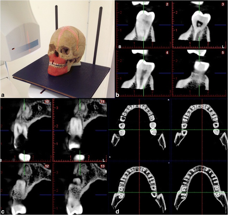
Methods: The study material comprised 12 dry skulls with maxilla and mandible. Artificial defects (dehiscence, tunnel, and fenestration) were created on anterior, premolar and molar teeth separately using burs. In total 14 dehiscences, 13 fenestrations, eight tunnel and 16 without periodontal defect were used in the study. These were randomly created on dry skulls. Each teeth with and without defects were images at various vertical angles using each of the following modalities: a Planmeca Promax Cone Beam CT and a Digora photostimulable phosphor plates. Specificity and sensitivity for assessing periodontal defects by each radiographic technique were calculated. Chi-square statistics were used to evaluate differences between modalities. Kappa statistics assessed the agreement between observers. Results were considered significant at P < 0.05.
Results: The kappa values for inter-observer agreement between observers ranged between 0.78 and 0.96 for the CBCT, and 0.43 and 0.72 of intraoral images. The Kappa values for detecting defects on anterior teeth was the least, following premolar and molar teeth both CBCT and intraoral imaging.
Conclusions: CBCT has the highest sensitivity and diagnostic accuracy for detecting various periodontal defects among the radiographic modalities examined.
Figures


References
Publication types
MeSH terms
LinkOut - more resources
Full Text Sources
Other Literature Sources
