

Evaluation of two novel ⁶⁴Cu-labeled RGD peptide radiotracers for enhanced PET imaging of tumor integrin αvβ₃
- PMID: 26016906
- PMCID: PMC4591102
- DOI: 10.1007/s00259-015-3085-7
Evaluation of two novel ⁶⁴Cu-labeled RGD peptide radiotracers for enhanced PET imaging of tumor integrin αvβ₃
Abstract
Purpose: Our goal was to demonstrate that suitably derivatized monomeric RGD peptide-based PET tracers, targeting integrin αvβ3, may offer advantages in image contrast, time for imaging, and low uptake in nontarget tissues.
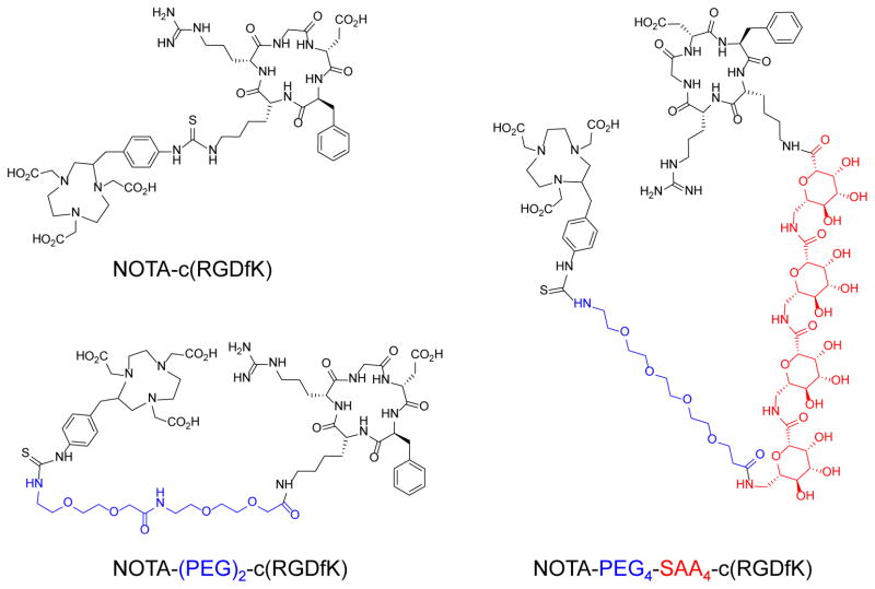
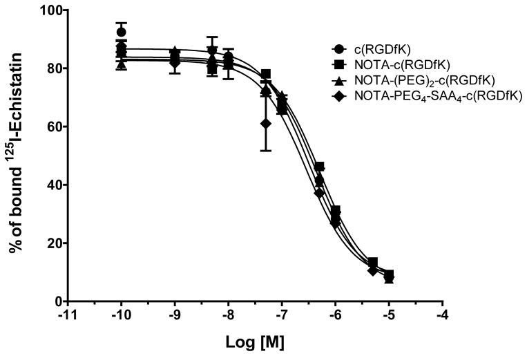
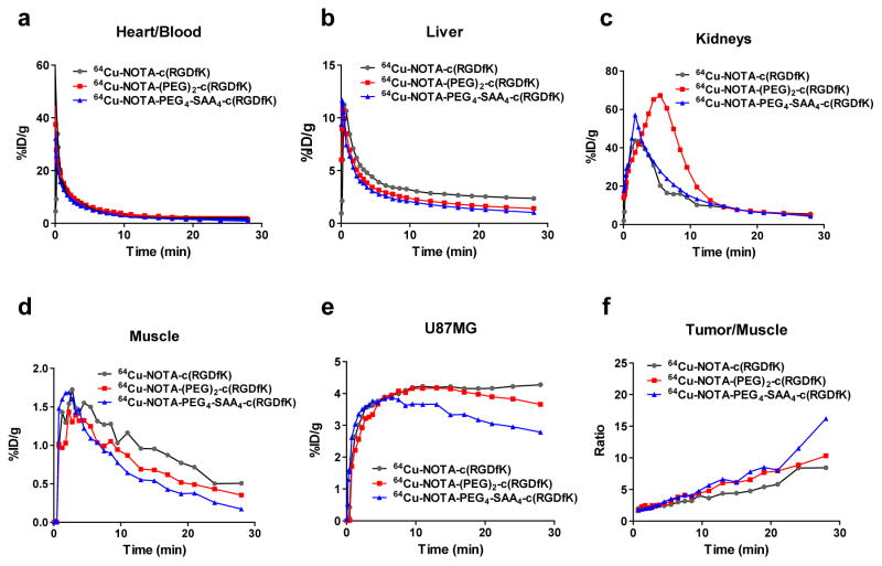
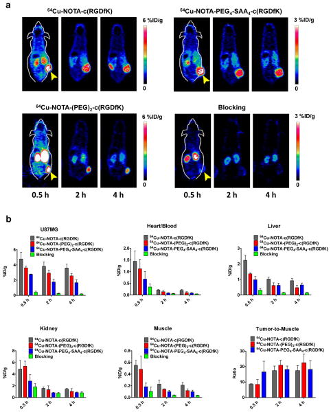
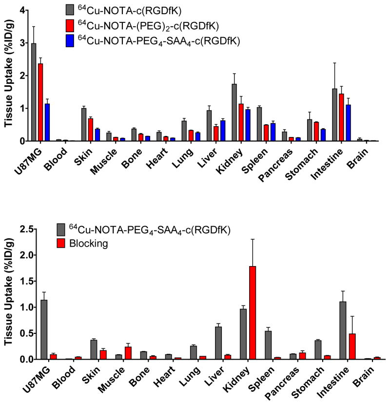
Methods: Two cyclic RGDfK derivatives, (PEG)2-c(RGDfK) and PEG4-SAA4-c(RGDfK), were constructed and conjugated to NOTA for (64)Cu labeling. Their integrin αvβ3-binding properties were determined via a competitive cell binding assay. Mice bearing U87MG tumors were intravenously injected with each of the (64)Cu-labeled peptides, and PET scans were acquired during the first 30 min, and 2 and 4 h after injection. Blocking and ex vivo biodistribution studies were carried out to validate the PET data and confirm the specificity of the tracers.
Results: The IC50 values of NOTA-(PEG)2-c(RGDfK) and NOTA-PEG4-SAA4-c(RGDfK) were 444 ± 41 nM and 288 ± 66 nM, respectively. Dynamic PET data of (64)Cu-NOTA-(PEG)2-c(RGDfK) and (64)Cu-NOTA-PEG4-SAA4-c(RGDfK) showed similar circulation t 1/2 and peak tumor uptake of about 4 %ID/g for both tracers. Due to its marked hydrophilicity, (64)Cu-NOTA-PEG4-SAA4-c(RGDfK) provided faster clearance from tumor and normal tissues yet maintained excellent tumor-to-background ratios. Static PET scans at later time-points corroborated the enhanced excretion of the tracer, especially from abdominal organs. Ex vivo biodistribution and receptor blocking studies confirmed the accuracy of the PET data and the integrin αvβ3-specificity of the peptides.
Conclusion: Our two novel RGD-based radiotracers with optimized pharmacokinetic properties allowed fast, high-contrast PET imaging of tumor-associated integrin αvβ3. These tracers may facilitate the imaging of abdominal malignancies, normally precluded by high background uptake.
Keywords: Angiogenesis; Copper-64 (64Cu); Integrin αvβ3; Molecular imaging; Positron emission tomography (PET); RGD peptide.
Conflict of interest statement
Figures
Similar articles
-
Pegylated Arg-Gly-Asp peptide: 64Cu labeling and PET imaging of brain tumor alphavbeta3-integrin expression.J Nucl Med. 2004 Oct;45(10):1776-83. J Nucl Med. 2004. PMID: 15471848
-
microPET imaging of glioma integrin {alpha}v{beta}3 expression using (64)Cu-labeled tetrameric RGD peptide.J Nucl Med. 2005 Oct;46(10):1707-18. J Nucl Med. 2005. PMID: 16204722
-
Novel (64)Cu- and (68)Ga-labeled RGD conjugates show improved PET imaging of α(ν)β(3) integrin expression and facile radiosynthesis.J Nucl Med. 2011 Aug;52(8):1276-84. doi: 10.2967/jnumed.111.087700. Epub 2011 Jul 15. J Nucl Med. 2011. PMID: 21764795
-
111In-Labeled 1,4,7,10-tetraazacyclododecane-1,4,7,10-tetracetic acid-Glu{PEG4-Glu[cyclo(Lys(Gly-Gly-Gly)-Arg-Gly-Asp-d-Phe)]-cyclo(Lys(Gly-Gly-Gly)-Arg-Gly-Asp-d-Phe)}-{PEG4-Glu[cyclo(Lys(Gly-Gly-Gly)-Arg-Gly-Asp-d-Phe)]-cyclo(Lys(Gly-Gly-Gly)-Arg-Gly-Asp-d-Phe)} (PEG4 = 15 amino-4,7,10,13-tetraoxapentadecanoic acid).2012 Feb 23 [updated 2012 Mar 22]. In: Molecular Imaging and Contrast Agent Database (MICAD) [Internet]. Bethesda (MD): National Center for Biotechnology Information (US); 2004–2013. 2012 Feb 23 [updated 2012 Mar 22]. In: Molecular Imaging and Contrast Agent Database (MICAD) [Internet]. Bethesda (MD): National Center for Biotechnology Information (US); 2004–2013. PMID: 22457888 Free Books & Documents. Review.
-
111In-Labeled 1,4,7,10-tetraazacyclododecane-1,4,7,10-tetracetic acid-Glu{PEG4-Glu[cyclo(Lys(PEG4)-Arg-Gly-Asp-d-Phe)]-cyclo(Lys(PEG4)-Arg-Gly-Asp-d-Phe)}-{PEG4-Glu[cyclo(Lys(PEG4)-Arg-Gly-Asp-d-Phe)]-cyclo(Lys(PEG4)-Arg-Gly-Asp-d-Phe)} (PEG4 = 15 amino-4,7,10,13-tetraoxapentadecanoic acid).2012 Feb 23 [updated 2012 Mar 22]. In: Molecular Imaging and Contrast Agent Database (MICAD) [Internet]. Bethesda (MD): National Center for Biotechnology Information (US); 2004–2013. 2012 Feb 23 [updated 2012 Mar 22]. In: Molecular Imaging and Contrast Agent Database (MICAD) [Internet]. Bethesda (MD): National Center for Biotechnology Information (US); 2004–2013. PMID: 22457890 Free Books & Documents. Review.
Cited by
-
Use of a leukocyte-targeted peptide probe as a potential tracer for imaging the tuberculosis granuloma.Tuberculosis (Edinb). 2018 Jan;108:201-210. doi: 10.1016/j.tube.2018.01.001. Epub 2018 Jan 8. Tuberculosis (Edinb). 2018. PMID: 29623875 Free PMC article.
-
A review of advances in the last decade on targeted cancer therapy using 177Lu: focusing on 177Lu produced by the direct neutron activation route.Am J Nucl Med Mol Imaging. 2021 Dec 15;11(6):443-475. eCollection 2021. Am J Nucl Med Mol Imaging. 2021. PMID: 35003885 Free PMC article. Review.
-
Mechanistic and quantitative insight into cell surface targeted molecular imaging agent design.Sci Rep. 2016 May 5;6:25424. doi: 10.1038/srep25424. Sci Rep. 2016. PMID: 27147293 Free PMC article.
-
The feasibility of 18F-AlF-NOTA-PRGD2 PET/CT for monitoring early response of Endostar antiangiogenic therapy in human nasopharyngeal carcinoma xenograft model compared with 18F-FDG.Oncotarget. 2016 May 10;7(19):27243-54. doi: 10.18632/oncotarget.8402. Oncotarget. 2016. PMID: 27029065 Free PMC article.
-
Multimodal imaging demonstrates enhanced tumor exposure of PEGylated FUD peptide in breast cancer.J Control Release. 2022 Oct;350:284-297. doi: 10.1016/j.jconrel.2022.08.028. Epub 2022 Aug 25. J Control Release. 2022. PMID: 35995299 Free PMC article.
References
-
- Hwang R, Varner J. The role of integrins in tumor angiogenesis. Hematol Oncol Clin North Am. 2004;18:991–1006. vii. - PubMed
-
- Robinson SD, Hodivala-Dilke KM. The role of beta3-integrins in tumor angiogenesis: context is everything. Curr Opin Cell Biol. 2011;23:630–7. - PubMed
-
- Puduvalli VK. Inhibition of angiogenesis as a therapeutic strategy against brain tumors. Cancer Treat Res. 2004;117:307–36. - PubMed
-
- Sengupta S, Chattopadhyay N, Mitra A, Ray S, Dasgupta S, Chatterjee A. Role of alphavbeta3 integrin receptors in breast tumor. J Exp Clin Cancer Res. 2001;20:585–90. - PubMed
-
- Felding-Habermann B, Fransvea E, O’Toole TE, Manzuk L, Faha B, Hensler M. Involvement of tumor cell integrin alpha v beta 3 in hematogenous metastasis of human melanoma cells. Clin Exp Metastasis. 2002;19:427–36. - PubMed
Publication types
MeSH terms
Substances
Grants and funding
LinkOut - more resources
Full Text Sources
Other Literature Sources
Miscellaneous
