. 2015 May 28:15:127.
doi: 10.1186/s12870-015-0525-2.

The UDP-glucose: glycoprotein glucosyltransferase (UGGT), a key enzyme in ER quality control, plays a significant role in plant growth as well as biotic and abiotic stress in Arabidopsis thaliana

Affiliations

The UDP-glucose: glycoprotein glucosyltransferase (UGGT), a key enzyme in ER quality control, plays a significant role in plant growth as well as biotic and abiotic stress in Arabidopsis thaliana

Francisca Blanco-Herrera et al. BMC Plant Biol. .

Abstract

Background: UDP-glucose: glycoprotein glucosyltransferase (UGGT) is a key player in the quality control mechanism (ER-QC) that newly synthesized glycoproteins undergo in the ER. It has been shown that the UGGT Arabidopsis orthologue is involved in ER-QC; however, its role in plant physiology remains unclear.

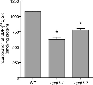
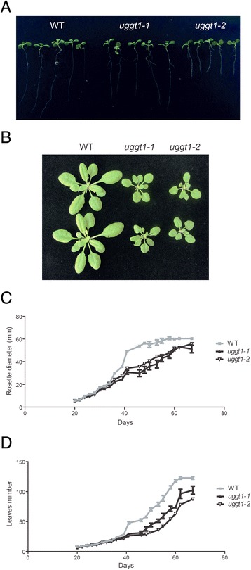
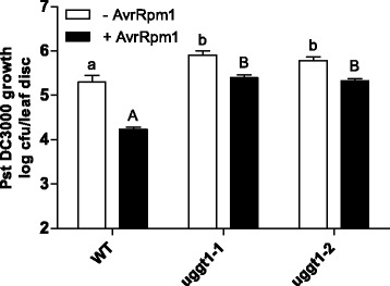
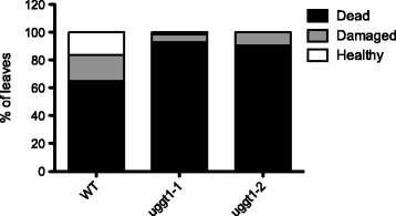
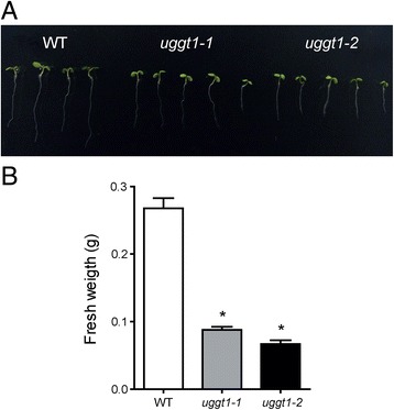
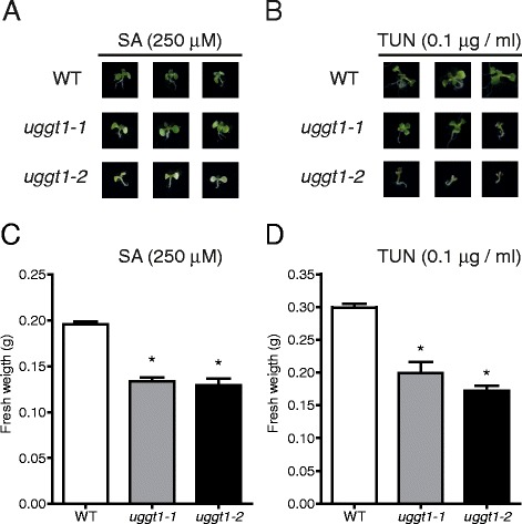
Results: Here, we show that two mutant alleles in the At1g71220 locus have none or reduced UGGT activity. In wild type plants, the AtUGGT transcript levels increased upon activation of the unfolded protein response (UPR). Interestingly, mutants in AtUGGT exhibited an endogenous up-regulation of genes that are UPR targets. In addition, mutants in AtUGGT showed a 30% reduction in the incorporation of UDP-Glucose into the ER suggesting that this enzyme drives the uptake of this substrate for the CNX/CRT cycle. Plants deficient in UGGT exhibited a delayed growth rate of the primary root and rosette as well as an alteration in the number of leaves. These mutants are more sensitive to pathogen attack as well as heat, salt, and UPR-inducing stressors. Additionally, the plants showed impairment in the establishment of systemic acquired resistance (SAR).

Conclusions: These results show that a lack of UGGT activity alters plant vegetative development and impairs the response to several abiotic and biotic stresses. Moreover, our results uncover an unexpected role of UGGT in the incorporation of UDP-Glucose into the ER lumen in Arabidopsis thaliana.


Figures

Fig. 1
Fig. 1
UGGT activity in ER-enriched fractions of Arabidopsis thaliana. a Microsomal membranes were prepared from etiolated Arabidopsis plants as described in the Material and Methods. The membranes were incubated in the presence of denatured SBA and UDP-[14C]glucose. The reaction was finished by adding TCA. The pellet was washed three times and the incorporated radioactivity was determined. Controls in which the SBA acceptor withheld or in which the ER-derived membranes were previously heat-inactivated are also shown. b The reaction was carried out as described above but with bovine thyroglobulin as the acceptor substrate. The proteins were treated with endoglycosidase H (Endo-H) to release the N-linked oligosaccharides that were separated by paper chromatography. The paper was cut and radioactivity determined by liquid scintillation
Fig. 2
Fig. 2
UGGT activity is reduced in plants bearing mutations in the At1g71220 gene. a UGGT activity detection in A. thaliana ER-enriched fractions from wild type and mutant plants. Incorporation of UDP-[14C]-glucose into unfolded SBA by wild type or mutant ER fractions. The upper panel shows the radioactivity associated with SBA while the lower panel shows the total amount of SBA used in the assay. b Quantification of the UGGT activity obtained from the PhosphorImager scans presented in a
Fig. 3
Fig. 3
UGGT is induced during UPR and UGGT mutants that exhibit ER stress. a Quantitative real-time PCR monitoring of AtUTr1, BiP1/2, BiP3, PDIL2-1 and AtUGGT transcript levels in stressed wild type plants. Fifteen day-old seedlings were treated over 5 hrs with DTT 2.5 mM or TUN 5 μg/ml in MS medium. Clathrin adapter (At5g46630) was used as a housekeeping gene. The average values of three independent experiments (n = 6) are shown; error bars represent ± SD. b Quantitative real-time PCR monitoring AtUTr1, BiP1/2, BiP3, PDIL2-1 and AtUGGT transcript levels in wild type and AtUGGT mutant plants grown under normal conditions. Fifteen day-old seedlings were used for the analysis. Clathrin adapter (At5g46630) was the housekeeping gene. The average values of three independent experiments (n = 6) are shown; error bars represent ± SD
Fig. 4
Fig. 4
The atuggt1-1 and atuggt1-2 mutants exhibit a reduced uptake of UDP-[14C]Glc into ER-derived vesicles. The UDP-Glc uptake was assayed into 50 μg of ER-enriched fractions from wild-type and AtUGGT mutant plants incubated with 1 μM UDP-[14C]Glc for 15 min. The reaction was stopped with a 10-fold dilution with cold STM buffer and filtration. The filter-associated label was counted using liquid scintillation. Results are presented as mean with SD; significance was determined with ANOVA. Asterisks indicate a Tukey’s test p-value < 0.001
Fig. 5
Fig. 5
UGGT mutant plants show altered growth rates during vegetative development compared to wild type. a Root length in seven day-old seedlings grown in half MS medium; both wild type and AtUGGT mutant plants are shown. b Phenotypes of six-week-old plants grown in hydroponic medium. The rosette diameter (c) and the number of leaves (d) were measured in plants between days 20 and 70. The average values of eight independent plants (n = 8) are shown; error bars represent ± SD
Fig. 6
Fig. 6
AtUGGT mutant plants are less tolerant to biotic stress. Whole leaves of four-week-old soil grown WT and mutant plants were infiltrated with Pst AvrRpm1 (OD600 = 0.001) to trigger SAR; a solution of 10 mM MgCl2 served as the mock. Twenty-four hours later the systemic leaves were infiltrated with Pst DC3000 (OD600 = 0.001). Bacterial growth (Pst DC3000) was monitored 3 days post infection. Error bars represent standard deviation from 6 samples. Different letters statically represent differences between the genotypes (lowercase for –AvrRpm1; uppercase for + AvrRpm1) at p < 0.05 (Tukey’s test). The experiments were performed at least three times with similar results
Fig. 7
Fig. 7
AtUGGT mutant plants are less tolerant to heat shock stress. Arabidopsis wild type and UGGT mutant plants were grown on soil for six weeks. The plants were treated at 42 °C for 2 hrs and returned to the growth chamber for 24 hrs. The leaves were then analyzed and classified as “dead” (completely dry and collapsed leaves), “damaged” (chlorotic lesions in leaves) or “healthy” (green and turgid leaves) and counted. The results are expressed as a percentage of total leaves analyzed per genotype (around 60 leaves per genotype)
Fig. 8
Fig. 8
AtUGGT mutant plants are less tolerant to salt stress. a Photograph of seven-day-old seedlings of the different genotypes grown in MS media supplemented with 150 mM NaCl. b Arabidopsis wild type or UGGT mutants were grown in MS media supplemented with 150 mM NaCl for 2 weeks. Eighty plants of each genotype were weighed, and the experiments were performed in triplicate. The average values of three independent plates (n = 240) are shown; error bars represent ± SD. Statistical significance was determined by ANOVA. Asterisks indicate a Tukey's test p-value <0.01
Fig. 9
Fig. 9
Mutant plants are over-sensitive to ER stress. Arabidopsis wild type and AtUGGT mutant plants were grown in MS media supplemented with 250 μM SA (a and c) or 0.1 μg/ml TUN (b) and (d) for 2 weeks. a and b. Photos of representative plants from different genotypes grown in SA or TUN, respectively. c and d. Eighty plants of each genotype were grown in SA or TUN, respectively, and were weighed in triplicates. The average values of three independent replicates (n = 240) are shown; error bars represent ± SD. Statistical significance was determined by ANOVA. Asterisks indicate a Tukey's test p-value < 0.01

Similar articles

Cited by

References

    1. Helenius A, Aebi M. Roles of N-linked glycans in the endoplasmic reticulum. Annu Rev Biochem. 2004;73:1019–49. doi: 10.1146/annurev.biochem.73.011303.073752. - DOI - PubMed
    1. Pattison RJ, Amtmann A. N-glycan production in the endoplasmic reticulum of plants. Trends Plant Sci. 2009;14:92–9. doi: 10.1016/j.tplants.2008.11.008. - DOI - PubMed
    1. Deprez P, Gautschi M, Helenius A. More than one glycan is needed for ER glucosidase II to allow entry of glycoproteins into the calnexin/calreticulin cycle. Mol Cell. 2005;19:183–95. doi: 10.1016/j.molcel.2005.05.029. - DOI - PubMed
    1. D’Alessio C, Caramelo JJ, Parodi AJ. UDP-Glc:glycoprotein glucosyltransferase-glucosidase II, the ying-yang of the ER quality control. Semin. Cell Dev Biol. 2010;21:491–9. doi: 10.1016/j.semcdb.2009.12.014. - DOI - PMC - PubMed
    1. Stigliano I, Alculumbre SG, Labriola CA, Parodi AJ, D’Alessio C. Glucosidase II and N-glycan mannose content regulate the half-lives of monoglucosylated species in vivo. Mol Biol Cell. 2011;22:1810–23. doi: 10.1091/mbc.E11-01-0019. - DOI - PMC - PubMed

Publication types

MeSH terms

Substances

LinkOut - more resources